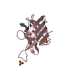
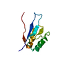
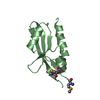
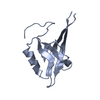
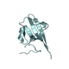
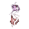
A: SH3 and multiple ankyrin repeat domains protein 3 B: SH3 and multiple ankyrin repeat domains protein 3 C: SH3 and multiple ankyrin repeat domains protein 3 D: SH3 and multiple ankyrin repeat domains protein 3 E: SH3 and multiple ankyrin repeat domains protein 3 F: SH3 and multiple ankyrin repeat domains protein 3 G: SH3 and multiple ankyrin repeat domains protein 3 H: SH3 and multiple ankyrin repeat domains protein 3 hetero molecules
Resolution: 1.83→1.88 Å / Rmerge(I) obs: 0.311 / Mean I/σ(I) obs: 2.36 / % possible all: 72.3
-
Processing
Software
Name
Version
Classification
MD2
datacollection
PHASES
phasing
REFMAC
5.5.0102
refinement
XDS
datareduction
XDS
datascaling
Refinement
Method to determine structure: MOLECULAR REPLACEMENT / Resolution: 1.83→33.97 Å / Cor.coef. Fo:Fc: 0.926 / Cor.coef. Fo:Fc free: 0.904 / SU B: 3.718 / SU ML: 0.11 / Cross valid method: THROUGHOUT / Stereochemistry target values: MAXIMUM LIKELIHOOD / Details: HYDROGENS HAVE BEEN ADDED IN THE RIDING POSITIONS
Rfactor
Num. reflection
% reflection
Selection details
Rfree
0.283
2852
5 %
RANDOM
Rwork
0.233
-
-
-
all
0.23562
56826
-
-
obs
0.23562
119285
88.73 %
-
Solvent computation
Ion probe radii: 0.8 Å / Shrinkage radii: 0.8 Å / VDW probe radii: 1.2 Å / Solvent model: MASK
Displacement parameters
Biso mean: 33.694 Å2
Baniso -1
Baniso -2
Baniso -3
1-
-21.29 Å2
0 Å2
-2.1 Å2
2-
-
29.47 Å2
0 Å2
3-
-
-
-8.18 Å2
Refinement step
Cycle: LAST / Resolution: 1.83→33.97 Å
Protein
Nucleic acid
Ligand
Solvent
Total
Num. atoms
6001
0
22
290
6313
Refine LS restraints
Refine-ID
Type
Dev ideal
Dev ideal target
Number
X-RAY DIFFRACTION
r_bond_refined_d
0.013
0.022
6160
X-RAY DIFFRACTION
r_angle_refined_deg
1.692
1.957
8331
X-RAY DIFFRACTION
r_dihedral_angle_1_deg
8.13
5
766
X-RAY DIFFRACTION
r_dihedral_angle_2_deg
36.98
23.227
282
X-RAY DIFFRACTION
r_dihedral_angle_3_deg
21.298
15
1051
X-RAY DIFFRACTION
r_dihedral_angle_4_deg
18.955
15
56
X-RAY DIFFRACTION
r_chiral_restr
0.117
0.2
960
X-RAY DIFFRACTION
r_gen_planes_refined
0.008
0.021
4611
X-RAY DIFFRACTION
r_mcbond_it
0.698
1.5
3807
X-RAY DIFFRACTION
r_mcangle_it
1.153
2
6146
X-RAY DIFFRACTION
r_scbond_it
1.764
3
2353
X-RAY DIFFRACTION
r_scangle_it
2.446
4.5
2180
+
About Yorodumi
-
News
-
Feb 9, 2022. New format data for meta-information of EMDB entries
New format data for meta-information of EMDB entries
Version 3 of the EMDB header file is now the official format.
The previous official version 1.9 will be removed from the archive.
In the structure databanks used in Yorodumi, some data are registered as the other names, "COVID-19 virus" and "2019-nCoV". Here are the details of the virus and the list of structure data.
Jan 31, 2019. EMDB accession codes are about to change! (news from PDBe EMDB page)
EMDB accession codes are about to change! (news from PDBe EMDB page)
The allocation of 4 digits for EMDB accession codes will soon come to an end. Whilst these codes will remain in use, new EMDB accession codes will include an additional digit and will expand incrementally as the available range of codes is exhausted. The current 4-digit format prefixed with “EMD-” (i.e. EMD-XXXX) will advance to a 5-digit format (i.e. EMD-XXXXX), and so on. It is currently estimated that the 4-digit codes will be depleted around Spring 2019, at which point the 5-digit format will come into force.
The EM Navigator/Yorodumi systems omit the EMD- prefix.
Related info.:Q: What is EMD? / ID/Accession-code notation in Yorodumi/EM Navigator
Yorodumi is a browser for structure data from EMDB, PDB, SASBDB, etc.
This page is also the successor to EM Navigator detail page, and also detail information page/front-end page for Omokage search.
The word "yorodu" (or yorozu) is an old Japanese word meaning "ten thousand". "mi" (miru) is to see.
Related info.:EMDB / PDB / SASBDB / Comparison of 3 databanks / Yorodumi Search / Aug 31, 2016. New EM Navigator & Yorodumi / Yorodumi Papers / Jmol/JSmol / Function and homology information / Changes in new EM Navigator and Yorodumi
 Movie
Movie Controller

 Yorodumi
 Open data
 Basic information
 Components
 Keywords
 Function and homology information
 X-RAY DIFFRACTION /
  Authors
 Citation
 Structure visualization
 Downloads & links
 Other downloads
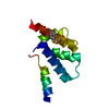


 PDBj
PDBj

 Assembly


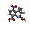
 Mass: 304.255 Da / Num. of mol.: 1 / Source method: obtained synthetically / Formula: C14H12N2O6
 Mass: 18.015 Da / Num. of mol.: 290 / Source method: isolated from a natural source / Formula: H2O
 Sample preparation
 / Beamline: 14.1 / Wavelength: 1.072 Å
 Processing