 Movie
Movie Controller

[English] 日本語
 Yorodumi
- PDB-3gxx: Structure of the SH2 domain of the Candida glabrata transcription... -
+ Open data

- Basic information
| Entry | Database: PDB / ID: 3gxx | ||||||
|---|---|---|---|---|---|---|---|
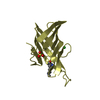
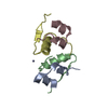
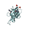
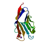
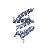
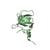
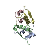
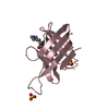
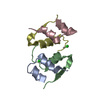
| Title | Structure of the SH2 domain of the Candida glabrata transcription elongation factor Spt6, crystal form B | ||||||
 Components Components | Transcription elongation factor SPT6 | ||||||
 Keywords Keywords | TRANSCRIPTION / SH2-fold / three stranded anti-parallel beta sheet / N-terminal alpha helix / C-terminal alpha helix / Nucleus / SH2 domain / Transcription regulation | ||||||
| Function / homology |  Function and homology information Function and homology informationtranscription antitermination factor activity, DNA binding / regulation of mRNA 3'-end processing / transcription elongation-coupled chromatin remodeling / poly(A)+ mRNA export from nucleus / cellular response to stress / pericentric heterochromatin / nucleosome binding / transcription elongation factor complex / positive regulation of transcription elongation by RNA polymerase II / G1/S transition of mitotic cell cycle ...transcription antitermination factor activity, DNA binding / regulation of mRNA 3'-end processing / transcription elongation-coupled chromatin remodeling / poly(A)+ mRNA export from nucleus / cellular response to stress / pericentric heterochromatin / nucleosome binding / transcription elongation factor complex / positive regulation of transcription elongation by RNA polymerase II / G1/S transition of mitotic cell cycle / euchromatin / nucleosome assembly / histone binding / negative regulation of transcription by RNA polymerase II / DNA binding Similarity search - Function | ||||||
| Biological species |  Candida glabrata (fungus) Candida glabrata (fungus) | ||||||
| Method |  X-RAY DIFFRACTION / SYNCHROTRON / MOLECULAR REPLACEMENT / Resolution: 2.4 Å X-RAY DIFFRACTION / SYNCHROTRON / MOLECULAR REPLACEMENT / Resolution: 2.4 Å | ||||||
 Authors Authors | Dengl, S. / Mayer, A. / Sun, M. / Cramer, P. | ||||||
 Citation Citation | Journal: J.Mol.Biol. / Year: 2009 Title: Structure and in vivo requirement of the yeast Spt6 SH2 domain Authors: Dengl, S. / Mayer, A. / Sun, M. / Cramer, P. | ||||||
| History |
|
- Structure visualization
| Structure viewer | Molecule: MolmilJmol/JSmol |
|---|
- Downloads & links
-Download
| PDBx/mmCIF format | 3gxx.cif.gz | 88.9 KB | Display | PDBx/mmCIF format |
|---|---|---|---|---|
| PDB format | pdb3gxx.ent.gz | 68.9 KB | Display | PDB format |
| PDBx/mmJSON format | 3gxx.json.gz | Tree view | PDBx/mmJSON format | |
| Others |  Other downloads Other downloads |
-Validation report
| Arichive directory | https://data.pdbj.org/pub/pdb/validation_reports/gx/3gxxftp://data.pdbj.org/pub/pdb/validation_reports/gx/3gxx | HTTPS FTP |
|---|
-Related structure data
| Related structure data |  3gxwSC S: Starting model for refinement C: citing same article ( |
|---|---|
| Similar structure data |

-Links
 PDBj
PDBj
- Assembly
| Deposited unit | 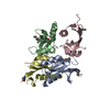
| ||||||||
|---|---|---|---|---|---|---|---|---|---|
| 1 | 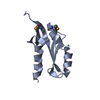
| ||||||||
| 2 | 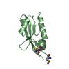
| ||||||||
| 3 | 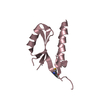
| ||||||||
| 4 | 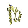
| ||||||||
| Unit cell |
|
-Components
| #1: Protein | Mass: 12426.574 Da / Num. of mol.: 4 / Fragment: SH2 domain / Mutation: L1309M, L1317M Source method: isolated from a genetically manipulated source Source: (gene. exp.) Candida glabrata (fungus) / Gene: CAGL0L04774g, SPT6 / Plasmid: pET28b / Production host:  #2: Water | ChemComp-HOH / |  Mass: 18.015 Da / Num. of mol.: 73 / Source method: isolated from a natural source / Formula: H2O Mass: 18.015 Da / Num. of mol.: 73 / Source method: isolated from a natural source / Formula: H2OHas protein modification | Y | |
|---|
-Experimental details
-Experiment
| Experiment | Method: X-RAY DIFFRACTION / Number of used crystals: 1 |
|---|
- Sample preparation
| Crystal | Density Matthews: 2.64 Å3/Da / Density % sol: 53.37 % |
|---|---|
| Crystal grow | Temperature: 293 K / Method: vapor diffusion, hanging drop / pH: 8 Details: 100 mM bicine pH 8.0, 4,3 M NaCl and 5 mM Tris(2-Carboxyethyl) phosphine Hydro-chloride, VAPOR DIFFUSION, HANGING DROP, temperature 293K |
-Data collection
| Diffraction | Mean temperature: 100 K |
|---|---|
| Diffraction source | Source: SYNCHROTRON / Site: BESSY  / Beamline: 14.1 / Wavelength: 0.97971 Å / Beamline: 14.1 / Wavelength: 0.97971 Å |
| Detector | Type: MARMOSAIC 225 mm CCD / Detector: CCD / Date: Aug 15, 2007 |
| Radiation | Protocol: SINGLE WAVELENGTH / Monochromatic (M) / Laue (L): M / Scattering type: x-ray |
| Radiation wavelength | Wavelength: 0.97971 Å / Relative weight: 1 |
| Reflection | Resolution: 2.4→20 Å / Num. all: 18513 / Num. obs: 18513 / % possible obs: 99.5 % / Observed criterion σ(F): 0 / Observed criterion σ(I): 0 / Redundancy: 3.9 % / Rsym value: 0.062 / Net I/σ(I): 45.1 |
| Reflection shell | Resolution: 2.4→2.49 Å / Redundancy: 3.7 % / Mean I/σ(I) obs: 7.7 / Rsym value: 0.19 / % possible all: 95.4 |
- Processing
| Software |
| ||||||||||||||||||||
|---|---|---|---|---|---|---|---|---|---|---|---|---|---|---|---|---|---|---|---|---|---|
| Refinement | Method to determine structure: MOLECULAR REPLACEMENT Starting model: PDB entry 3GXW Resolution: 2.4→18.7 Å / Stereochemistry target values: Engh & Huber
| ||||||||||||||||||||
| Refinement step | Cycle: LAST / Resolution: 2.4→18.7 Å
|
