 Movie
Movie Controller

[English] 日本語
 Yorodumi
- PDB-2g9n: Structure of the DEAD domain of Human eukaryotic initiation facto... -
+ Open data

- Basic information
| Entry | Database: PDB / ID: 2g9n | ||||||
|---|---|---|---|---|---|---|---|
| Title | Structure of the DEAD domain of Human eukaryotic initiation factor 4A, eIF4A | ||||||
 Components Components | Eukaryotic initiation factor 4A-I | ||||||
 Keywords Keywords | HYDROLASE / DEAD-box / Helicase / DDX2A / RNA / Structural Genomics / Structural Genomics Consortium / SGC | ||||||
| Function / homology |  Function and homology information Function and homology informationActivation of the mRNA upon binding of the cap-binding complex and eIFs, and subsequent binding to 43S / RNA cap binding / eukaryotic translation initiation factor 4F complex / nuclear stress granule / Z-decay: degradation of maternal mRNAs by zygotically expressed factors / translation factor activity, RNA binding / Deadenylation of mRNA / M-decay: degradation of maternal mRNAs by maternally stored factors / cytoplasmic translational initiation / Ribosomal scanning and start codon recognition ...Activation of the mRNA upon binding of the cap-binding complex and eIFs, and subsequent binding to 43S / RNA cap binding / eukaryotic translation initiation factor 4F complex / nuclear stress granule / Z-decay: degradation of maternal mRNAs by zygotically expressed factors / translation factor activity, RNA binding / Deadenylation of mRNA / M-decay: degradation of maternal mRNAs by maternally stored factors / cytoplasmic translational initiation / Ribosomal scanning and start codon recognition / Translation initiation complex formation / Dengue Virus Genome Translation and Replication / GTP hydrolysis and joining of the 60S ribosomal subunit / L13a-mediated translational silencing of Ceruloplasmin expression / translation initiation factor activity / helicase activity / translational initiation / ISG15 antiviral mechanism / cytoplasmic stress granule / double-stranded RNA binding / RNA helicase activity / RNA helicase / mRNA binding / perinuclear region of cytoplasm / positive regulation of transcription by RNA polymerase II / ATP hydrolysis activity / RNA binding / extracellular exosome / ATP binding / membrane / plasma membrane / cytosol / cytoplasm Similarity search - Function | ||||||
| Biological species |  Homo sapiens (human) Homo sapiens (human) | ||||||
| Method |  X-RAY DIFFRACTION / SYNCHROTRON / MOLECULAR REPLACEMENT / Resolution: 2.25 Å X-RAY DIFFRACTION / SYNCHROTRON / MOLECULAR REPLACEMENT / Resolution: 2.25 Å | ||||||
 Authors Authors | Hogbom, M. / Ogg, D. / Arrowsmith, C. / Berglund, H. / Collins, R. / Edwards, A. / Ehn, M. / Flodin, S. / Flores, A. / Graslund, S. ...Hogbom, M. / Ogg, D. / Arrowsmith, C. / Berglund, H. / Collins, R. / Edwards, A. / Ehn, M. / Flodin, S. / Flores, A. / Graslund, S. / Hallberg, B.M. / Hammarstrom, M. / Kotenyova, T. / Nilsson-Ehle, P. / Nordlund, P. / Nyman, T. / Persson, C. / Sagemark, J. / Stenmark, P. / Sundstrom, M. / Thorsell, A.G. / Uppenberg, J. / Van Den Berg, S. / Weigelt, J. / Holmberg-Schiavone, L. / Structural Genomics Consortium (SGC) | ||||||
 Citation Citation | Journal: Plos One / Year: 2010 Title: Comparative Structural Analysis of Human DEAD-Box RNA Helicases. Authors: Schutz, P. / Karlberg, T. / van den Berg, S. / Collins, R. / Lehtio, L. / Hogbom, M. / Holmberg-Schiavone, L. / Tempel, W. / Park, H.W. / Hammarstrom, M. / Moche, M. / Thorsell, A.G. / Schuler, H. | ||||||
| History |
|
- Structure visualization
| Structure viewer | Molecule: MolmilJmol/JSmol |
|---|
- Downloads & links
-Download
| PDBx/mmCIF format | 2g9n.cif.gz | 104.5 KB | Display | PDBx/mmCIF format |
|---|---|---|---|---|
| PDB format | pdb2g9n.ent.gz | 81.9 KB | Display | PDB format |
| PDBx/mmJSON format | 2g9n.json.gz | Tree view | PDBx/mmJSON format | |
| Others |  Other downloads Other downloads |
-Validation report
| Arichive directory | https://data.pdbj.org/pub/pdb/validation_reports/g9/2g9nftp://data.pdbj.org/pub/pdb/validation_reports/g9/2g9n | HTTPS FTP |
|---|
-Related structure data
| Related structure data |  2p6nC  2pl3C  2rb4C  3b7gC  3berC  3borC  3dkpC  3fe2C  3iuyC  3ly5C 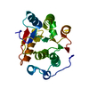 1qdeS S: Starting model for refinement C: citing same article ( |
|---|---|
| Similar structure data |


-Links
 PDBj
PDBj


- Assembly
| Deposited unit | 
| |||||||||||||||||||||||||||||||||||||||||||||||||||||||||||||||||
|---|---|---|---|---|---|---|---|---|---|---|---|---|---|---|---|---|---|---|---|---|---|---|---|---|---|---|---|---|---|---|---|---|---|---|---|---|---|---|---|---|---|---|---|---|---|---|---|---|---|---|---|---|---|---|---|---|---|---|---|---|---|---|---|---|---|---|
| 1 | 
| |||||||||||||||||||||||||||||||||||||||||||||||||||||||||||||||||
| 2 | 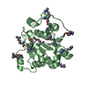
| |||||||||||||||||||||||||||||||||||||||||||||||||||||||||||||||||
| Unit cell |
| |||||||||||||||||||||||||||||||||||||||||||||||||||||||||||||||||
| Noncrystallographic symmetry (NCS) | NCS domain:
NCS domain segments: Ens-ID: 1 / Refine code: 4
|
-Components
| #1: Protein | Mass: 25087.316 Da / Num. of mol.: 2 / Fragment: DEAD domain Source method: isolated from a genetically manipulated source Source: (gene. exp.) Homo sapiens (human) / Gene: EIF4A1, DDX2A, EIF4A / Plasmid: pNIC-28-Bsa1 / Production host:  References: UniProt: P60842, Hydrolases; Acting on acid anhydrides; In phosphorus-containing anhydrides #2: Water | ChemComp-HOH / |  Mass: 18.015 Da / Num. of mol.: 226 / Source method: isolated from a natural source / Formula: H2O Mass: 18.015 Da / Num. of mol.: 226 / Source method: isolated from a natural source / Formula: H2O |
|---|
-Experimental details
-Experiment
| Experiment | Method: X-RAY DIFFRACTION / Number of used crystals: 1 |
|---|
- Sample preparation
| Crystal | Density Matthews: 2.14 Å3/Da / Density % sol: 42.56 % |
|---|---|
| Crystal grow | Temperature: 293 K / Method: vapor diffusion, sitting drop / pH: 7.5 Details: 20% PEG3350, 200 mM Ammonium Nitrate, 500 mM NaCl, pH 7.5, VAPOR DIFFUSION, SITTING DROP, temperature 293K |
-Data collection
| Diffraction source | Source: SYNCHROTRON / Site: ESRF  / Beamline: ID14-2 / Wavelength: 0.933 Å / Beamline: ID14-2 / Wavelength: 0.933 Å |
|---|---|
| Detector | Type: ADSC QUANTUM 4 / Detector: CCD / Date: Feb 12, 2006 |
| Radiation | Monochromator: Diamond (111), Ge(220) / Protocol: SINGLE WAVELENGTH / Monochromatic (M) / Laue (L): M / Scattering type: x-ray |
| Radiation wavelength | Wavelength: 0.933 Å / Relative weight: 1 |
| Reflection | Resolution: 2.25→20 Å / Num. all: 19100 / Num. obs: 19100 / % possible obs: 99.65 % / Observed criterion σ(F): 0 / Observed criterion σ(I): 0 / Redundancy: 3.45 % / Biso Wilson estimate: 25.9 Å2 / Rsym value: 0.071 / Net I/σ(I): 7.4 |
| Reflection shell | Resolution: 2.25→2.37 Å / Redundancy: 3.46 % / Mean I/σ(I) obs: 5.3 / Num. unique all: 2912 / Rsym value: 0.241 / % possible all: 100 |
- Processing
| Software |
| ||||||||||||||||||||||||||||||||||||||||||||||||||||||||||||||||||||||||||||||||||||||||||
|---|---|---|---|---|---|---|---|---|---|---|---|---|---|---|---|---|---|---|---|---|---|---|---|---|---|---|---|---|---|---|---|---|---|---|---|---|---|---|---|---|---|---|---|---|---|---|---|---|---|---|---|---|---|---|---|---|---|---|---|---|---|---|---|---|---|---|---|---|---|---|---|---|---|---|---|---|---|---|---|---|---|---|---|---|---|---|---|---|---|---|---|
| Refinement | Method to determine structure: MOLECULAR REPLACEMENT Starting model: pdb entry 1QDE Resolution: 2.25→20 Å / Cor.coef. Fo:Fc: 0.942 / Cor.coef. Fo:Fc free: 0.882 / SU B: 12.622 / SU ML: 0.166 / Cross valid method: THROUGHOUT / σ(F): 0 / ESU R: 0.365 / ESU R Free: 0.258 / Stereochemistry target values: MAXIMUM LIKELIHOOD / Details: HYDROGENS HAVE BEEN ADDED IN THE RIDING POSITIONS
| ||||||||||||||||||||||||||||||||||||||||||||||||||||||||||||||||||||||||||||||||||||||||||
| Solvent computation | Ion probe radii: 0.8 Å / Shrinkage radii: 0.8 Å / VDW probe radii: 1.4 Å / Solvent model: MASK | ||||||||||||||||||||||||||||||||||||||||||||||||||||||||||||||||||||||||||||||||||||||||||
| Displacement parameters | Biso mean: 14.89 Å2
| ||||||||||||||||||||||||||||||||||||||||||||||||||||||||||||||||||||||||||||||||||||||||||
| Refinement step | Cycle: LAST / Resolution: 2.25→20 Å
| ||||||||||||||||||||||||||||||||||||||||||||||||||||||||||||||||||||||||||||||||||||||||||
| Refine LS restraints |
| ||||||||||||||||||||||||||||||||||||||||||||||||||||||||||||||||||||||||||||||||||||||||||
| Refine LS restraints NCS | Dom-ID: 1 / Auth asym-ID: A / Ens-ID: 1 / Number: 1514 / Refine-ID: X-RAY DIFFRACTION
| ||||||||||||||||||||||||||||||||||||||||||||||||||||||||||||||||||||||||||||||||||||||||||
| LS refinement shell | Resolution: 2.25→2.308 Å / Total num. of bins used: 20
|
