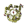
 Movie
Movie Controller


+ Open data

- Basic information
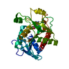
| Entry | Database: PDB / ID: 3l9c | ||||||
|---|---|---|---|---|---|---|---|
| Title | The Crystal Structure of smu.777 from Streptococcus mutans UA159 | ||||||
 Components Components | 3-dehydroquinate dehydratase | ||||||
 Keywords Keywords | LYASE / aroD / Amino-acid biosynthesis / Aromatic amino acid biosynthesis / Schiff base | ||||||
| Function / homology |  Function and homology information Function and homology information3,4-dihydroxybenzoate biosynthetic process / 3-dehydroquinate dehydratase / 3-dehydroquinate dehydratase activity / chorismate biosynthetic process / aromatic amino acid biosynthetic process / amino acid biosynthetic process Similarity search - Function | ||||||
| Biological species |  Streptococcus mutans (bacteria) Streptococcus mutans (bacteria) | ||||||
| Method |  X-RAY DIFFRACTION / SYNCHROTRON / MOLECULAR REPLACEMENT / Resolution: 1.6 Å X-RAY DIFFRACTION / SYNCHROTRON / MOLECULAR REPLACEMENT / Resolution: 1.6 Å | ||||||
 Authors Authors | Su, X.-D. / Huang, Y.H. / Liu, X. | ||||||
 Citation Citation | Journal: TO BE PUBLISHED Title: The Crystal Structure of smu.777 from Streptococcus mutans UA159 Authors: Su, X.-D. / Huang, Y.H. / Liu, X. | ||||||
| History |
|
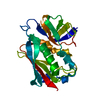
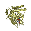
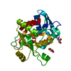
- Structure visualization
| Structure viewer | Molecule: MolmilJmol/JSmol |
|---|
- Downloads & links
-Download
| PDBx/mmCIF format | 3l9c.cif.gz | 103.1 KB | Display | PDBx/mmCIF format |
|---|---|---|---|---|
| PDB format | pdb3l9c.ent.gz | 78.2 KB | Display | PDB format |
| PDBx/mmJSON format | 3l9c.json.gz | Tree view | PDBx/mmJSON format | |
| Others |  Other downloads Other downloads |
-Validation report
| Arichive directory | https://data.pdbj.org/pub/pdb/validation_reports/l9/3l9cftp://data.pdbj.org/pub/pdb/validation_reports/l9/3l9c | HTTPS FTP |
|---|
-Related structure data
| Related structure data |  2oczS S: Starting model for refinement |
|---|---|
| Similar structure data |


-Links
 PDBj
PDBj
- Assembly
| Deposited unit | 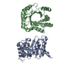
| ||||||||
|---|---|---|---|---|---|---|---|---|---|
| 1 | 
| ||||||||
| 2 | 
| ||||||||
| Unit cell |
|
-Components
| #1: Protein | Mass: 29358.191 Da / Num. of mol.: 2 Source method: isolated from a genetically manipulated source Source: (gene. exp.) Streptococcus mutans (bacteria) / Strain: UA159 / Gene: smu.777 / Plasmid: pET28a / Production host: #2: Water | ChemComp-HOH / |  Mass: 18.015 Da / Num. of mol.: 145 / Source method: isolated from a natural source / Formula: H2O Mass: 18.015 Da / Num. of mol.: 145 / Source method: isolated from a natural source / Formula: H2O |
|---|
-Experimental details
-Experiment
| Experiment | Method: X-RAY DIFFRACTION / Number of used crystals: 1 |
|---|
- Sample preparation
| Crystal | Density Matthews: 1.93 Å3/Da / Density % sol: 36.16 % |
|---|---|
| Crystal grow | Temperature: 289 K / Method: vapor diffusion, sitting drop / pH: 7.5 Details: 0.05M Magnesium Chloride hexahydrate, 0.1M HEPES pH7.5, 30% PEGMME550, 0.01M Phenol, VAPOR DIFFUSION, SITTING DROP, temperature 289K |
-Data collection
| Diffraction | Mean temperature: 100 K |
|---|---|
| Diffraction source | Source: SYNCHROTRON / Site: SSRF  / Beamline: BL17U / Wavelength: 0.97156 Å / Beamline: BL17U / Wavelength: 0.97156 Å |
| Detector | Type: MARMOSAIC 225 mm CCD / Detector: CCD / Date: May 9, 2009 |
| Radiation | Protocol: SINGLE WAVELENGTH / Monochromatic (M) / Laue (L): M / Scattering type: x-ray |
| Radiation wavelength | Wavelength: 0.97156 Å / Relative weight: 1 |
| Reflection | Resolution: 1.54→30 Å / Num. all: 66520 / Num. obs: 66453 / % possible obs: 99.9 % / Observed criterion σ(I): 1 / Biso Wilson estimate: 20.77 Å2 |
| Reflection shell | Resolution: 1.54→1.56 Å / % possible all: 100 |
- Processing
| Software |
| |||||||||||||||||||||||||||||||||||||||||||||||||||||||||||||||||||||||||||||||||||||||||||||||||||||||||||||||||||||||||||||||||||||||||||||||||||||||||||||||||
|---|---|---|---|---|---|---|---|---|---|---|---|---|---|---|---|---|---|---|---|---|---|---|---|---|---|---|---|---|---|---|---|---|---|---|---|---|---|---|---|---|---|---|---|---|---|---|---|---|---|---|---|---|---|---|---|---|---|---|---|---|---|---|---|---|---|---|---|---|---|---|---|---|---|---|---|---|---|---|---|---|---|---|---|---|---|---|---|---|---|---|---|---|---|---|---|---|---|---|---|---|---|---|---|---|---|---|---|---|---|---|---|---|---|---|---|---|---|---|---|---|---|---|---|---|---|---|---|---|---|---|---|---|---|---|---|---|---|---|---|---|---|---|---|---|---|---|---|---|---|---|---|---|---|---|---|---|---|---|---|---|---|---|
| Refinement | Method to determine structure: MOLECULAR REPLACEMENT Starting model: PDB ENTRY 2ocz Resolution: 1.6→28.888 Å / Occupancy max: 1 / Occupancy min: 0.5 / FOM work R set: 0.788 / SU ML: 0.23 / σ(F): 1.89 / Phase error: 28 / Stereochemistry target values: ML
| |||||||||||||||||||||||||||||||||||||||||||||||||||||||||||||||||||||||||||||||||||||||||||||||||||||||||||||||||||||||||||||||||||||||||||||||||||||||||||||||||
| Solvent computation | Shrinkage radii: 0.9 Å / VDW probe radii: 1.11 Å / Solvent model: FLAT BULK SOLVENT MODEL / Bsol: 50.293 Å2 / ksol: 0.377 e/Å3 | |||||||||||||||||||||||||||||||||||||||||||||||||||||||||||||||||||||||||||||||||||||||||||||||||||||||||||||||||||||||||||||||||||||||||||||||||||||||||||||||||
| Displacement parameters | Biso max: 80.89 Å2 / Biso mean: 27.333 Å2 / Biso min: 12.41 Å2
| |||||||||||||||||||||||||||||||||||||||||||||||||||||||||||||||||||||||||||||||||||||||||||||||||||||||||||||||||||||||||||||||||||||||||||||||||||||||||||||||||
| Refinement step | Cycle: LAST / Resolution: 1.6→28.888 Å
| |||||||||||||||||||||||||||||||||||||||||||||||||||||||||||||||||||||||||||||||||||||||||||||||||||||||||||||||||||||||||||||||||||||||||||||||||||||||||||||||||
| Refine LS restraints |
| |||||||||||||||||||||||||||||||||||||||||||||||||||||||||||||||||||||||||||||||||||||||||||||||||||||||||||||||||||||||||||||||||||||||||||||||||||||||||||||||||
| LS refinement shell |
|
