 Movie
Movie Controller

[English] 日本語
 Yorodumi
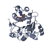
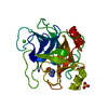
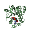
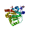
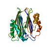
- PDB-1qde: CRYSTAL STRUCTURE OF THE ATPASE DOMAIN OF TRANSLATION INITIATION ... -
+ Open data

- Basic information
| Entry | Database: PDB / ID: 1qde | ||||||
|---|---|---|---|---|---|---|---|
| Title | CRYSTAL STRUCTURE OF THE ATPASE DOMAIN OF TRANSLATION INITIATION FACTOR 4A FROM SACCHAROMYCES CEREVISIAE-THE PROTOTYPE OF THE DEAD BOX PROTEIN FAMILY | ||||||
 Components Components | TRANSLATION INITIATION FACTOR 4A | ||||||
 Keywords Keywords | GENE REGULATION / TRANSLATION INITIATION / SACCHAROMYCES CEREVISIAE / DEAD BOX PROTEIN FAMILY | ||||||
| Function / homology |  Function and homology information Function and homology informationActivation of the mRNA upon binding of the cap-binding complex and eIFs, and subsequent binding to 43S / positive regulation of formation of translation preinitiation complex / Deadenylation of mRNA / eukaryotic translation initiation factor 4F complex / ATP-dependent activity, acting on RNA / regulation of translational initiation / Translation initiation complex formation / Ribosomal scanning and start codon recognition / cytoplasmic translational initiation / L13a-mediated translational silencing of Ceruloplasmin expression ...Activation of the mRNA upon binding of the cap-binding complex and eIFs, and subsequent binding to 43S / positive regulation of formation of translation preinitiation complex / Deadenylation of mRNA / eukaryotic translation initiation factor 4F complex / ATP-dependent activity, acting on RNA / regulation of translational initiation / Translation initiation complex formation / Ribosomal scanning and start codon recognition / cytoplasmic translational initiation / L13a-mediated translational silencing of Ceruloplasmin expression / translation initiation factor activity / translational initiation / cytoplasmic stress granule / RNA helicase activity / RNA helicase / ribosome / ATP hydrolysis activity / RNA binding / ATP binding / plasma membrane / cytoplasm Similarity search - Function | ||||||
| Biological species |  | ||||||
| Method |  X-RAY DIFFRACTION / Resolution: 2 Å X-RAY DIFFRACTION / Resolution: 2 Å | ||||||
 Authors Authors | Benz, J. / Trachsel, H. / Baumann, U. | ||||||
 Citation Citation | Journal: Structure Fold.Des. / Year: 1999 Title: Crystal structure of the ATPase domain of translation initiation factor 4A from Saccharomyces cerevisiae--the prototype of the DEAD box protein family. Authors: Benz, J. / Trachsel, H. / Baumann, U. | ||||||
| History |
|
- Structure visualization
| Structure viewer | Molecule: MolmilJmol/JSmol |
|---|
- Downloads & links
-Download
| PDBx/mmCIF format | 1qde.cif.gz | 51.6 KB | Display | PDBx/mmCIF format |
|---|---|---|---|---|
| PDB format | pdb1qde.ent.gz | 37.5 KB | Display | PDB format |
| PDBx/mmJSON format | 1qde.json.gz | Tree view | PDBx/mmJSON format | |
| Others |  Other downloads Other downloads |
-Validation report
| Arichive directory | https://data.pdbj.org/pub/pdb/validation_reports/qd/1qdeftp://data.pdbj.org/pub/pdb/validation_reports/qd/1qde | HTTPS FTP |
|---|
-Related structure data
| Similar structure data |
|---|


-Links
 PDBj
PDBj

- Assembly
| Deposited unit | 
| ||||||||
|---|---|---|---|---|---|---|---|---|---|
| 1 |
| ||||||||
| Unit cell |
|
-Components
| #1: Protein | Mass: 25165.982 Da / Num. of mol.: 1 / Fragment: N-TERMINAL DOMAIN, RESIDUES 9-232 Source method: isolated from a genetically manipulated source Source: (gene. exp.) Plasmid: PQE30 / Production host:  |
|---|---|
| #2: Chemical | ChemComp-SO4 /   Mass: 96.063 Da / Num. of mol.: 1 / Source method: obtained synthetically / Formula: SO4 Mass: 96.063 Da / Num. of mol.: 1 / Source method: obtained synthetically / Formula: SO4 |
| #3: Water | ChemComp-HOH /  Mass: 18.015 Da / Num. of mol.: 50 / Source method: isolated from a natural source / Formula: H2O Mass: 18.015 Da / Num. of mol.: 50 / Source method: isolated from a natural source / Formula: H2O |
-Experimental details
-Experiment
| Experiment | Method: X-RAY DIFFRACTION / Number of used crystals: 1 |
|---|
- Sample preparation
| Crystal | Density Matthews: 2.11 Å3/Da / Density % sol: 41.62 % | ||||||||||||||||||||||||||||||||||||||||||
|---|---|---|---|---|---|---|---|---|---|---|---|---|---|---|---|---|---|---|---|---|---|---|---|---|---|---|---|---|---|---|---|---|---|---|---|---|---|---|---|---|---|---|---|
| Crystal grow | Temperature: 293 K / Method: vapor diffusion, sitting drop / pH: 4.8 Details: PEG 8000, MGCL2, (NH4)2SO4, MES, pH 4.8, VAPOR DIFFUSION, SITTING DROP, temperature 293K | ||||||||||||||||||||||||||||||||||||||||||
| Crystal | *PLUS Density % sol: 48 % | ||||||||||||||||||||||||||||||||||||||||||
| Crystal grow | *PLUS Temperature: 20 ℃ / pH: 8.8 / Method: vapor diffusion, hanging drop | ||||||||||||||||||||||||||||||||||||||||||
| Components of the solutions | *PLUS
|
-Data collection
| Diffraction | Mean temperature: 293 K |
|---|---|
| Diffraction source | Source: ROTATING ANODE / Type: RIGAKU RU200 / Wavelength: 1.5418 |
| Detector | Type: RIGAKU RAXIS IV / Detector: IMAGE PLATE |
| Radiation | Protocol: SINGLE WAVELENGTH / Monochromatic (M) / Laue (L): M / Scattering type: x-ray |
| Radiation wavelength | Wavelength: 1.5418 Å / Relative weight: 1 |
| Reflection | Resolution: 2→40 Å / Num. all: 14988 / Num. obs: 14988 / % possible obs: 99.6 % / Observed criterion σ(I): 0 / Redundancy: 6.07 % / Biso Wilson estimate: 22.4 Å2 / Rmerge(I) obs: 0.09 / Net I/σ(I): 14.5 |
| Reflection shell | Resolution: 2→2.03 Å / Redundancy: 6.4 % / Rmerge(I) obs: 0.315 / % possible all: 99.1 |
| Reflection | *PLUS Rmerge(I) obs: 0.09 |
- Processing
| Software |
| ||||||||||||||||||||||||||||||||||||||||||||||||||||||||||||
|---|---|---|---|---|---|---|---|---|---|---|---|---|---|---|---|---|---|---|---|---|---|---|---|---|---|---|---|---|---|---|---|---|---|---|---|---|---|---|---|---|---|---|---|---|---|---|---|---|---|---|---|---|---|---|---|---|---|---|---|---|---|
| Refinement | Resolution: 2→40 Å / σ(F): 0 / σ(I): 0 / Stereochemistry target values: ENGH & HUBER
| ||||||||||||||||||||||||||||||||||||||||||||||||||||||||||||
| Refinement step | Cycle: LAST / Resolution: 2→40 Å
| ||||||||||||||||||||||||||||||||||||||||||||||||||||||||||||
| Refine LS restraints |
| ||||||||||||||||||||||||||||||||||||||||||||||||||||||||||||
| Software | *PLUS Name: CNS / Classification: refinement | ||||||||||||||||||||||||||||||||||||||||||||||||||||||||||||
| Refinement | *PLUS Highest resolution: 2 Å / Lowest resolution: 40 Å / σ(F): 0 / % reflection Rfree: 10 % / Rfactor obs: 0.213 | ||||||||||||||||||||||||||||||||||||||||||||||||||||||||||||
| Solvent computation | *PLUS | ||||||||||||||||||||||||||||||||||||||||||||||||||||||||||||
| Displacement parameters | *PLUS | ||||||||||||||||||||||||||||||||||||||||||||||||||||||||||||
| Refine LS restraints | *PLUS Type: c_angle_deg / Dev ideal: 1.4 |
