 Movie
Movie Controller

[English] 日本語
 Yorodumi
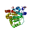
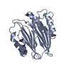
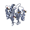
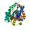
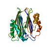
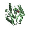
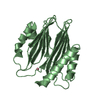
- PDB-3w42: Crystal structure of RsbX in complex with manganese in space group P1 -
+ Open data

- Basic information
| Entry | Database: PDB / ID: 3w42 | ||||||
|---|---|---|---|---|---|---|---|
| Title | Crystal structure of RsbX in complex with manganese in space group P1 | ||||||
 Components Components | Phosphoserine phosphatase RsbX | ||||||
 Keywords Keywords | HYDROLASE / Signaling protein / Stressosome / Environmental stress / Phosphoric Monoester Hydrolases / Dephosphorylation / Tertiary / phosphatase / Protein phosphatase / magnesium/manganese binding / alpha-beta beta-alpha sandwich fold | ||||||
| Function / homology |  Function and homology information Function and homology informationphosphoserine phosphatase / L-phosphoserine phosphatase activity / phosphoprotein phosphatase activity / response to heat Similarity search - Function | ||||||
| Biological species |  | ||||||
| Method |  X-RAY DIFFRACTION / SYNCHROTRON / MOLECULAR REPLACEMENT / Resolution: 1.06 Å X-RAY DIFFRACTION / SYNCHROTRON / MOLECULAR REPLACEMENT / Resolution: 1.06 Å | ||||||
 Authors Authors | Teh, A.H. / Makino, M. / Baba, S. / Shimizu, N. / Yamamoto, M. / Kumasaka, T. | ||||||
 Citation Citation | Journal: Acta Crystallogr.,Sect.D / Year: 2015 Title: Structure of the RsbX phosphatase involved in the general stress response of Bacillus subtilis Authors: Teh, A.H. / Makino, M. / Hoshino, T. / Baba, S. / Shimizu, N. / Yamamoto, M. / Kumasaka, T. | ||||||
| History |
|
- Structure visualization
| Structure viewer | Molecule: MolmilJmol/JSmol |
|---|
- Downloads & links
-Download
| PDBx/mmCIF format | 3w42.cif.gz | 206.3 KB | Display | PDBx/mmCIF format |
|---|---|---|---|---|
| PDB format | pdb3w42.ent.gz | 165.4 KB | Display | PDB format |
| PDBx/mmJSON format | 3w42.json.gz | Tree view | PDBx/mmJSON format | |
| Others |  Other downloads Other downloads |
-Validation report
| Arichive directory | https://data.pdbj.org/pub/pdb/validation_reports/w4/3w42ftp://data.pdbj.org/pub/pdb/validation_reports/w4/3w42 | HTTPS FTP |
|---|
-Related structure data
| Related structure data | 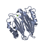 3w40C  3w41C  3w43C  3w44C  3w45SC C: citing same article ( S: Starting model for refinement |
|---|---|
| Similar structure data |


-Links
 PDBj
PDBj- Assembly
| Deposited unit | 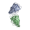
| ||||||||
|---|---|---|---|---|---|---|---|---|---|
| 1 | 
| ||||||||
| 2 | 
| ||||||||
| Unit cell |
|
-Components
| #1: Protein | Mass: 22172.029 Da / Num. of mol.: 2 Source method: isolated from a genetically manipulated source Source: (gene. exp.) #2: Chemical |   Mass: 54.938 Da / Num. of mol.: 2 / Source method: obtained synthetically / Formula: Mn Mass: 54.938 Da / Num. of mol.: 2 / Source method: obtained synthetically / Formula: Mn#3: Water | ChemComp-HOH / |  Mass: 18.015 Da / Num. of mol.: 609 / Source method: isolated from a natural source / Formula: H2O Mass: 18.015 Da / Num. of mol.: 609 / Source method: isolated from a natural source / Formula: H2O |
|---|
-Experimental details
-Experiment
| Experiment | Method: X-RAY DIFFRACTION / Number of used crystals: 1 |
|---|
- Sample preparation
| Crystal | Density Matthews: 2.01 Å3/Da / Density % sol: 38.68 % |
|---|---|
| Crystal grow | Temperature: 298 K / Method: vapor diffusion / pH: 8.5 Details: 20 % PEG 1000, 0.1M Tris-HCl, 5mM MgCl2, 3mM MnCl2, pH 8.5, VAPOR DIFFUSION, temperature 298K |
-Data collection
| Diffraction | Mean temperature: 100 K |
|---|---|
| Diffraction source | Source: SYNCHROTRON / Site: SPring-8  / Beamline: BL41XU / Wavelength: 1 Å / Beamline: BL41XU / Wavelength: 1 Å |
| Detector | Type: MARMOSAIC 225 mm CCD / Detector: CCD / Date: Feb 18, 2009 / Details: KIRKPATRICK-BAEZ MIRRORS |
| Radiation | Monochromator: Si(111) / Protocol: SINGLE WAVELENGTH / Monochromatic (M) / Laue (L): M / Scattering type: x-ray |
| Radiation wavelength | Wavelength: 1 Å / Relative weight: 1 |
| Reflection | Resolution: 1.06→50 Å / Num. obs: 155995 / % possible obs: 90.3 % / Observed criterion σ(I): -3 / Redundancy: 3.6 % / Rsym value: 0.081 / Net I/σ(I): 23.6 |
| Reflection shell | Resolution: 1.06→1.1 Å / Redundancy: 2.5 % / Mean I/σ(I) obs: 2.3 / Rsym value: 0.366 / % possible all: 71.5 |
- Processing
| Software |
| ||||||||||||||||||||||||||||||||||||||||||||||||||||||||||||||||||||||||||||||||||||||||||||||||||||||||||||||||||||||||||||||||||||||||||||||||||||||||||||||||||||||||||
|---|---|---|---|---|---|---|---|---|---|---|---|---|---|---|---|---|---|---|---|---|---|---|---|---|---|---|---|---|---|---|---|---|---|---|---|---|---|---|---|---|---|---|---|---|---|---|---|---|---|---|---|---|---|---|---|---|---|---|---|---|---|---|---|---|---|---|---|---|---|---|---|---|---|---|---|---|---|---|---|---|---|---|---|---|---|---|---|---|---|---|---|---|---|---|---|---|---|---|---|---|---|---|---|---|---|---|---|---|---|---|---|---|---|---|---|---|---|---|---|---|---|---|---|---|---|---|---|---|---|---|---|---|---|---|---|---|---|---|---|---|---|---|---|---|---|---|---|---|---|---|---|---|---|---|---|---|---|---|---|---|---|---|---|---|---|---|---|---|---|---|---|
| Refinement | Method to determine structure: MOLECULAR REPLACEMENT Starting model: PDB ENTRY 3W45 Resolution: 1.06→20 Å / Cor.coef. Fo:Fc: 0.977 / Cor.coef. Fo:Fc free: 0.969 / Occupancy max: 1 / Occupancy min: 0.02 / SU B: 0.756 / SU ML: 0.018 / SU R Cruickshank DPI: 0.0284 / Cross valid method: THROUGHOUT / ESU R: 0.028 / ESU R Free: 0.03 / Stereochemistry target values: MAXIMUM LIKELIHOOD / Details: HYDROGENS HAVE BEEN ADDED IN THE RIDING POSITIONS
| ||||||||||||||||||||||||||||||||||||||||||||||||||||||||||||||||||||||||||||||||||||||||||||||||||||||||||||||||||||||||||||||||||||||||||||||||||||||||||||||||||||||||||
| Solvent computation | Ion probe radii: 0.8 Å / Shrinkage radii: 0.8 Å / VDW probe radii: 1.2 Å / Solvent model: BABINET MODEL WITH MASK | ||||||||||||||||||||||||||||||||||||||||||||||||||||||||||||||||||||||||||||||||||||||||||||||||||||||||||||||||||||||||||||||||||||||||||||||||||||||||||||||||||||||||||
| Displacement parameters | Biso mean: 8.484 Å2
| ||||||||||||||||||||||||||||||||||||||||||||||||||||||||||||||||||||||||||||||||||||||||||||||||||||||||||||||||||||||||||||||||||||||||||||||||||||||||||||||||||||||||||
| Refinement step | Cycle: LAST / Resolution: 1.06→20 Å
| ||||||||||||||||||||||||||||||||||||||||||||||||||||||||||||||||||||||||||||||||||||||||||||||||||||||||||||||||||||||||||||||||||||||||||||||||||||||||||||||||||||||||||
| Refine LS restraints |
| ||||||||||||||||||||||||||||||||||||||||||||||||||||||||||||||||||||||||||||||||||||||||||||||||||||||||||||||||||||||||||||||||||||||||||||||||||||||||||||||||||||||||||
| LS refinement shell | Resolution: 1.061→1.088 Å / Total num. of bins used: 20
|
