deNEDDylase activity / protein deubiquitination / post-translational protein modification / protein catabolic process / Synthesis of active ubiquitin: roles of E1 and E2 enzymes / ubiquitin binding / UCH proteinases / peptidase activity / Neddylation / ubiquitin-dependent protein catabolic process ...deNEDDylase activity / protein deubiquitination / post-translational protein modification / protein catabolic process / Synthesis of active ubiquitin: roles of E1 and E2 enzymes / ubiquitin binding / UCH proteinases / peptidase activity / Neddylation / ubiquitin-dependent protein catabolic process / cysteine-type deubiquitinase activity / ubiquitinyl hydrolase 1 / protein ubiquitination / Golgi apparatus / nucleoplasm / cytosol / cytoplasm Similarity search - Function
Mass: 18.015 Da / Num. of mol.: 104 / Source method: isolated from a natural source / Formula: H2O
-
Experimental details
-
Experiment
Experiment
Method: X-RAY DIFFRACTION / Number of used crystals: 1
-
Sample preparation
Crystal
Density Matthews: 2.37 Å3/Da / Density % sol: 48 %
Crystal grow
Temperature: 277 K / Method: vapor diffusion, sitting drop / pH: 6.7 Details: PROTEIN AT 12 MG/ML WAS CRYSTALLIZED FROM 26%(W/W) PEG 4000, 200 MM SODIUM ACETATE, 100 MM PIPES PH 6.7 AND 10 MM DTT IN SITTING WELLS AT 4 DEGREES CELSIUS., vapor diffusion - sitting drop, temperature 277K
Crystal grow
*PLUS
Temperature: 4 ℃ / Method: vapor diffusion, sitting drop
Monochromatic (M) / Laue (L): M / Scattering type: x-ray
Radiation wavelength
Wavelength: 1.08 Å / Relative weight: 1
Reflection
Resolution: 1.8→30 Å / Num. obs: 23334 / % possible obs: 98 % / Observed criterion σ(I): -2 / Redundancy: 4.5 % / Biso Wilson estimate: 20.7 Å2 / Rmerge(I) obs: 0.04 / Rsym value: 0.04 / Net I/σ(I): 20
Reflection shell
Resolution: 1.8→1.83 Å / Redundancy: 3 % / Rmerge(I) obs: 0.19 / Mean I/σ(I) obs: 5 / Rsym value: 0.19 / % possible all: 93
Reflection shell
*PLUS
% possible obs: 93 %
-
Processing
Software
Name
Version
Classification
X-PLOR
3.843
modelbuilding
X-PLOR
3.843
refinement
DENZO
datareduction
SCALEPACK
datascaling
X-PLOR
3.843
phasing
Refinement
Method to determine structure: MAD / Resolution: 1.8→6 Å / Rfactor Rfree error: 0.009 / Data cutoff high absF: 10000000 / Data cutoff low absF: 0.001 / Isotropic thermal model: RESTRAINED / Cross valid method: THROUGHOUT / σ(F): 0 Details: THERE IS A DISORDERED REGION BETWEEN RESIDUES 146 AND 167.
Rfactor
Num. reflection
% reflection
Selection details
Rfree
0.294
1056
4.8 %
RANDOM
Rwork
0.232
-
-
-
obs
0.232
21819
97.7 %
-
Displacement parameters
Biso mean: 28.4 Å2
Refine analyze
Free
Obs
Luzzati coordinate error
0.31 Å
0.25 Å
Luzzati d res low
-
5 Å
Luzzati sigma a
0.25 Å
0.28 Å
Refinement step
Cycle: LAST / Resolution: 1.8→6 Å
Protein
Nucleic acid
Ligand
Solvent
Total
Num. atoms
1661
0
0
312
1973
Refine LS restraints
Refine-ID
Type
Dev ideal
Dev ideal target
X-RAY DIFFRACTION
x_bond_d
0.01
X-RAY DIFFRACTION
x_bond_d_na
X-RAY DIFFRACTION
x_bond_d_prot
X-RAY DIFFRACTION
x_angle_d
X-RAY DIFFRACTION
x_angle_d_na
X-RAY DIFFRACTION
x_angle_d_prot
X-RAY DIFFRACTION
x_angle_deg
1.1
X-RAY DIFFRACTION
x_angle_deg_na
X-RAY DIFFRACTION
x_angle_deg_prot
X-RAY DIFFRACTION
x_dihedral_angle_d
25
X-RAY DIFFRACTION
x_dihedral_angle_d_na
X-RAY DIFFRACTION
x_dihedral_angle_d_prot
X-RAY DIFFRACTION
x_improper_angle_d
1.98
X-RAY DIFFRACTION
x_improper_angle_d_na
X-RAY DIFFRACTION
x_improper_angle_d_prot
X-RAY DIFFRACTION
x_mcbond_it
2.17
0.75
X-RAY DIFFRACTION
x_mcangle_it
3.29
1
X-RAY DIFFRACTION
x_scbond_it
3.73
1
X-RAY DIFFRACTION
x_scangle_it
5.67
1.25
LS refinement shell
Resolution: 1.8→1.91 Å / Rfactor Rfree error: 0.029 / Total num. of bins used: 6
In the structure databanks used in Yorodumi, some data are registered as the other names, "COVID-19 virus" and "2019-nCoV". Here are the details of the virus and the list of structure data.
Jan 31, 2019. EMDB accession codes are about to change! (news from PDBe EMDB page)
EMDB accession codes are about to change! (news from PDBe EMDB page)
The allocation of 4 digits for EMDB accession codes will soon come to an end. Whilst these codes will remain in use, new EMDB accession codes will include an additional digit and will expand incrementally as the available range of codes is exhausted. The current 4-digit format prefixed with “EMD-” (i.e. EMD-XXXX) will advance to a 5-digit format (i.e. EMD-XXXXX), and so on. It is currently estimated that the 4-digit codes will be depleted around Spring 2019, at which point the 5-digit format will come into force.
The EM Navigator/Yorodumi systems omit the EMD- prefix.
Related info.:Q: What is EMD? / ID/Accession-code notation in Yorodumi/EM Navigator
Yorodumi is a browser for structure data from EMDB, PDB, SASBDB, etc.
This page is also the successor to EM Navigator detail page, and also detail information page/front-end page for Omokage search.
The word "yorodu" (or yorozu) is an old Japanese word meaning "ten thousand". "mi" (miru) is to see.
Related info.:EMDB / PDB / SASBDB / Comparison of 3 databanks / Yorodumi Search / Aug 31, 2016. New EM Navigator & Yorodumi / Yorodumi Papers / Jmol/JSmol / Function and homology information / Changes in new EM Navigator and Yorodumi
 Movie
Movie Controller

 Yorodumi
 Open data
 Basic information
 Components
 Keywords
 Function and homology information
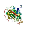
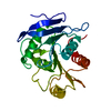
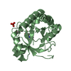
 Homo sapiens (human)
 X-RAY DIFFRACTION /
  Authors
 Citation
 Structure visualization
 Downloads & links
 Other downloads
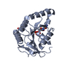


 PDBj
PDBj

 Assembly

 Mass: 18.015 Da / Num. of mol.: 104 / Source method: isolated from a natural source / Formula: H2O
 Sample preparation
 / Beamline: BL7-1 / Wavelength: 1.08
  Processing