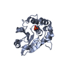
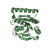
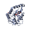
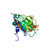
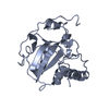
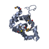
- PDB-6ent: Structure of the rat RKIP variant delta143-146 -
+
Open data
ID or keywords:
Loading...
-
Basic information
Entry
Database: PDB / ID: 6ent
Title
Structure of the rat RKIP variant delta143-146
Components
Phosphatidylethanolamine-binding protein 1
Keywords
SIGNALING PROTEIN / RKIP / PEBP / PBP
Function / homology
Function and homology information
positive regulation of acetylcholine metabolic process / positive regulation of acetylcholine biosynthetic process / Negative regulation of MAPK pathway / MAP2K and MAPK activation / regulation of the force of heart contraction / receptor serine/threonine kinase binding / mitogen-activated protein kinase binding / sperm capacitation / response to corticosterone / eating behavior ...positive regulation of acetylcholine metabolic process / positive regulation of acetylcholine biosynthetic process / Negative regulation of MAPK pathway / MAP2K and MAPK activation / regulation of the force of heart contraction / receptor serine/threonine kinase binding / mitogen-activated protein kinase binding / sperm capacitation / response to corticosterone / eating behavior / spermatid development / negative regulation of protein phosphorylation / positive regulation of cAMP/PKA signal transduction / response to electrical stimulus / response to cAMP / negative regulation of MAPK cascade / positive regulation of mitotic nuclear division / axon terminus / response to activity / hippocampus development / serine-type endopeptidase inhibitor activity / response to calcium ion / response to toxic substance / kinase binding / apical part of cell / MAPK cascade / synaptic vesicle / response to oxidative stress / response to ethanol / mitochondrial outer membrane / response to xenobiotic stimulus / signaling receptor binding / neuronal cell body / lipid binding / protein kinase binding / enzyme binding / cell surface / : / ATP binding Similarity search - Function
Phosphatidylethanolamine-binding, conserved site / Phosphatidylethanolamine-binding protein family signature. / Phosphatidylethanolamine-binding Protein / PEBP-like / Phosphatidylethanolamine-binding protein, eukaryotic / Phosphatidylethanolamine-binding protein / Phosphatidylethanolamine-binding protein / PEBP-like superfamily / Alpha-Beta Complex / Alpha Beta Similarity search - Domain/homology
Resolution: 2.66→20 Å / Cor.coef. Fo:Fc: 0.952 / Cor.coef. Fo:Fc free: 0.933 / SU B: 31.629 / SU ML: 0.293 / Cross valid method: THROUGHOUT / ESU R: 0.849 / ESU R Free: 0.322 / Details: HYDROGENS HAVE BEEN ADDED IN THE RIDING POSITIONS
Rfactor
Num. reflection
% reflection
Selection details
Rfree
0.25043
315
4.7 %
RANDOM
Rwork
0.20959
-
-
-
obs
0.21148
6365
99.66 %
-
Solvent computation
Ion probe radii: 0.8 Å / Shrinkage radii: 0.8 Å / VDW probe radii: 1.2 Å
 Movie
Movie Controller


 Open data
 Basic information
 Components
 Keywords
 Function and homology information
 X-RAY DIFFRACTION /
  Authors
 United States,
  Germany, 3items
  Citation
 Structure visualization
 Downloads & links
 Other downloads
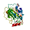


 PDBj
PDBj Assembly
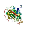


 Mass: 65.409 Da / Num. of mol.: 1 / Source method: obtained synthetically / Formula: Zn
 Mass: 35.453 Da / Num. of mol.: 1 / Source method: obtained synthetically / Formula: Cl
 Mass: 18.015 Da / Num. of mol.: 6 / Source method: isolated from a natural source / Formula: H2O
 Sample preparation
 Processing