 Movie
Movie Controller


+ Open data

- Basic information
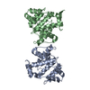
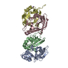
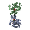
| Entry | Database: PDB / ID: 1bqy | ||||||
|---|---|---|---|---|---|---|---|
| Title | Plasminogen activator (TSV-PA) from snake venom | ||||||
 Components Components | PLASMINOGEN ACTIVATOR | ||||||
 Keywords Keywords | HYDROLASE/HYDROLASE INHIBITOR / FIBRINOLYSIS / PLASMINOGEN ACTIVATOR / SERINE PROTEINASE / SNAKE VENOM / HYDROLASE-HYDROLASE INHIBITOR COMPLEX / BLOOD CLOTTING | ||||||
| Function / homology |  Function and homology information Function and homology informationHydrolases; Acting on peptide bonds (peptidases); Serine endopeptidases / secretory granule / toxin activity / serine-type endopeptidase activity / proteolysis / extracellular region Similarity search - Function | ||||||
| Biological species |  Viridovipera stejnegeri (Stejneger's pit viper) Viridovipera stejnegeri (Stejneger's pit viper) | ||||||
| Method |  X-RAY DIFFRACTION / MOLECULAR REPLACEMENT / Resolution: 2.5 Å X-RAY DIFFRACTION / MOLECULAR REPLACEMENT / Resolution: 2.5 Å | ||||||
 Authors Authors | Parry, M.A.A. / Bode, W. | ||||||
 Citation Citation | Journal: Structure / Year: 1998 Title: The crystal structure of the novel snake venom plasminogen activator TSV-PA: a prototype structure for snake venom serine proteinases. Authors: Parry, M.A. / Jacob, U. / Huber, R. / Wisner, A. / Bon, C. / Bode, W. | ||||||
| History |
|
- Structure visualization
| Structure viewer | Molecule: MolmilJmol/JSmol |
|---|
- Downloads & links
-Download
| PDBx/mmCIF format | 1bqy.cif.gz | 107 KB | Display | PDBx/mmCIF format |
|---|---|---|---|---|
| PDB format | pdb1bqy.ent.gz | 82 KB | Display | PDB format |
| PDBx/mmJSON format | 1bqy.json.gz | Tree view | PDBx/mmJSON format | |
| Others |  Other downloads Other downloads |
-Validation report
| Arichive directory | https://data.pdbj.org/pub/pdb/validation_reports/bq/1bqyftp://data.pdbj.org/pub/pdb/validation_reports/bq/1bqy | HTTPS FTP |
|---|
-Related structure data
| Related structure data | 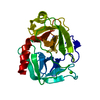 3ptbS S: Starting model for refinement |
|---|---|
| Similar structure data |


-Links
 PDBj
PDBj- Assembly
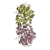
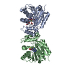
| Deposited unit | 
| ||||||||
|---|---|---|---|---|---|---|---|---|---|
| 1 | 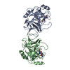
| ||||||||
| 2 | 
| ||||||||
| 3 | 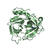
| ||||||||
| Unit cell |
|
-Components
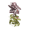
| #1: Protein | Mass: 25634.023 Da / Num. of mol.: 2 Source method: isolated from a genetically manipulated source Source: (gene. exp.) Viridovipera stejnegeri (Stejneger's pit viper)Organ: VENOM GLAND / Plasmid: PET / Production host:  #2: Chemical | 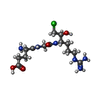  Type: peptide-like, Peptide-like / Class: Inhibitor / Mass: 395.862 Da / Num. of mol.: 2 / Source method: obtained synthetically / Formula: C14H28ClN6O5 Type: peptide-like, Peptide-like / Class: Inhibitor / Mass: 395.862 Da / Num. of mol.: 2 / Source method: obtained synthetically / Formula: C14H28ClN6O5Details: CHLOROMETHYLKETONE INHIBITOR COVALENTLY BOUND TO THE ACTIVE SITE SER 195 AND HIS 57 References: GLU-GLY-ARG-CHLOROMETHYL KETONE #3: Water | ChemComp-HOH / |  Mass: 18.015 Da / Num. of mol.: 162 / Source method: isolated from a natural source / Formula: H2O Mass: 18.015 Da / Num. of mol.: 162 / Source method: isolated from a natural source / Formula: H2OHas protein modification | Y | Nonpolymer details | THE UNBOUND FORM OF THE INHIBITOR IS GLU-GLY-ARG-CMK-CHLOROMETHYLKETONE. UPON REACTION WITH PROTEIN ...THE UNBOUND FORM OF THE INHIBITOR IS GLU-GLY-ARG-CMK-CHLOROMETH | |
|---|
-Experimental details
-Experiment
| Experiment | Method: X-RAY DIFFRACTION / Number of used crystals: 1 |
|---|
- Sample preparation
| Crystal | Density Matthews: 2.6 Å3/Da / Density % sol: 50 % | ||||||||||||||||||||||||||||||
|---|---|---|---|---|---|---|---|---|---|---|---|---|---|---|---|---|---|---|---|---|---|---|---|---|---|---|---|---|---|---|---|
| Crystal grow | *PLUS pH: 4.3 / Method: vapor diffusion, sitting drop | ||||||||||||||||||||||||||||||
| Components of the solutions | *PLUS
|
-Data collection
| Diffraction | Mean temperature: 293 K |
|---|---|
| Diffraction source | Wavelength: 1.5418 |
| Detector | Type: MARRESEARCH / Detector: IMAGE PLATE / Date: Apr 1, 1997 / Details: MIRRORS |
| Radiation | Monochromator: CU FILTER / Protocol: SINGLE WAVELENGTH / Monochromatic (M) / Laue (L): M / Scattering type: x-ray |
| Radiation wavelength | Wavelength: 1.5418 Å / Relative weight: 1 |
| Reflection | Resolution: 2.5→30 Å / Num. obs: 17988 / % possible obs: 97.7 % / Observed criterion σ(I): 2 / Redundancy: 2 % / Rmerge(I) obs: 0.102 |
| Reflection shell | Resolution: 2.5→2.59 Å / Rmerge(I) obs: 0.444 / % possible all: 82.1 |
| Reflection | *PLUS Num. measured all: 91942 |
| Reflection shell | *PLUS % possible obs: 82.1 % |
- Processing
| Software |
| ||||||||||||||||||||||||||||||||||||||||||||||||||||||||||||
|---|---|---|---|---|---|---|---|---|---|---|---|---|---|---|---|---|---|---|---|---|---|---|---|---|---|---|---|---|---|---|---|---|---|---|---|---|---|---|---|---|---|---|---|---|---|---|---|---|---|---|---|---|---|---|---|---|---|---|---|---|---|
| Refinement | Method to determine structure: MOLECULAR REPLACEMENT Starting model: PDB ENTRY 3PTB Resolution: 2.5→30 Å / Data cutoff high absF: 10000000 / Data cutoff low absF: 0.001 / σ(F): 2
| ||||||||||||||||||||||||||||||||||||||||||||||||||||||||||||
| Refinement step | Cycle: LAST / Resolution: 2.5→30 Å
| ||||||||||||||||||||||||||||||||||||||||||||||||||||||||||||
| Refine LS restraints |
| ||||||||||||||||||||||||||||||||||||||||||||||||||||||||||||
| Refine LS restraints NCS | NCS model details: RESTRAINTS | ||||||||||||||||||||||||||||||||||||||||||||||||||||||||||||
| Software | *PLUS Name: X-PLOR / Version: 3.8 / Classification: refinement | ||||||||||||||||||||||||||||||||||||||||||||||||||||||||||||
| LS refinement shell | *PLUS Highest resolution: 2.5 Å / Lowest resolution: 2.59 Å / Rfactor Rwork: 0.352 |
