 Movie
Movie Controller


+ Open data

- Basic information
| Entry | Database: PDB / ID: 1oa8 | ||||||
|---|---|---|---|---|---|---|---|
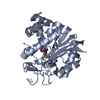
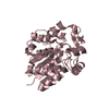
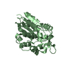
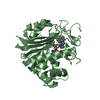
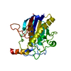
| Title | AXH domain of human spinocerebellar ataxin-1 | ||||||
 Components Components | ATAXIN-1 | ||||||
 Keywords Keywords | RNA BINDING / HIGH MOBILITY GROUP HOMOLOGY / HMG / RNA-BINDING / DIMERIZATION | ||||||
| Function / homology |  Function and homology information Function and homology informationpoly(G) binding / negative regulation of insulin-like growth factor receptor signaling pathway / POZ domain binding / nuclear inclusion body / nuclear export / lung alveolus development / poly(U) RNA binding / positive regulation of glial cell proliferation / social behavior / RNA processing ...poly(G) binding / negative regulation of insulin-like growth factor receptor signaling pathway / POZ domain binding / nuclear inclusion body / nuclear export / lung alveolus development / poly(U) RNA binding / positive regulation of glial cell proliferation / social behavior / RNA processing / adult locomotory behavior / insulin-like growth factor receptor signaling pathway / excitatory postsynaptic potential / learning / brain development / visual learning / memory / nuclear matrix / transcription by RNA polymerase II / postsynapse / negative regulation of DNA-templated transcription / chromatin binding / nucleolus / negative regulation of transcription by RNA polymerase II / positive regulation of transcription by RNA polymerase II / protein-containing complex / DNA binding / RNA binding / nucleoplasm / identical protein binding / nucleus / cytosol / cytoplasm Similarity search - Function | ||||||
| Biological species |  HOMO SAPIENS (human) HOMO SAPIENS (human) | ||||||
| Method |  X-RAY DIFFRACTION / SYNCHROTRON / MAD / Resolution: 1.7 Å X-RAY DIFFRACTION / SYNCHROTRON / MAD / Resolution: 1.7 Å | ||||||
 Authors Authors | Allen, M.D. / Chen, Y.W. / Bycroft, M. | ||||||
 Citation Citation | Journal: J. Biol. Chem. / Year: 2004 Title: The structure of the AXH domain of spinocerebellar ataxin-1. Authors: Chen, Y.W. / Allen, M.D. / Veprintsev, D.B. / Lowe, J. / Bycroft, M. #1: Journal: FEBS Lett. / Year: 2003 Title: The Axh Module: An Independently Folded Domain Common to Ataxin-1 and Hbp1. Authors: De Chiara, C. / Giannini, C. / Adinolfi, S. / De Boer, J. / Guida, S. / Ramos, A. / Jodice, C. / Kioussis, D. / Pastore, A. | ||||||
| History |
|
- Structure visualization
| Structure viewer | Molecule: MolmilJmol/JSmol |
|---|
- Downloads & links
-Download
| PDBx/mmCIF format | 1oa8.cif.gz | 117.4 KB | Display | PDBx/mmCIF format |
|---|---|---|---|---|
| PDB format | pdb1oa8.ent.gz | 91.5 KB | Display | PDB format |
| PDBx/mmJSON format | 1oa8.json.gz | Tree view | PDBx/mmJSON format | |
| Others |  Other downloads Other downloads |
-Validation report
| Arichive directory | https://data.pdbj.org/pub/pdb/validation_reports/oa/1oa8ftp://data.pdbj.org/pub/pdb/validation_reports/oa/1oa8 | HTTPS FTP |
|---|
-Related structure data
| Similar structure data |
|---|


-Links
 PDBj
PDBj- Assembly
| Deposited unit | 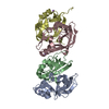
| ||||||||||||||||
|---|---|---|---|---|---|---|---|---|---|---|---|---|---|---|---|---|---|
| 1 | 
| ||||||||||||||||
| 2 | 
| ||||||||||||||||
| Unit cell |
| ||||||||||||||||
| Noncrystallographic symmetry (NCS) | NCS oper:
|
-Components
| #1: Protein | Mass: 14428.339 Da / Num. of mol.: 4 / Fragment: AXH DOMAIN, RESIDUES 562-694 Source method: isolated from a genetically manipulated source Source: (gene. exp.) HOMO SAPIENS (human) / Plasmid: PRSETA / Production host:  #2: Chemical |   Mass: 22.990 Da / Num. of mol.: 3 / Source method: obtained synthetically / Formula: Na Mass: 22.990 Da / Num. of mol.: 3 / Source method: obtained synthetically / Formula: Na#3: Water | ChemComp-HOH / |  Mass: 18.015 Da / Num. of mol.: 378 / Source method: isolated from a natural source / Formula: H2O Mass: 18.015 Da / Num. of mol.: 378 / Source method: isolated from a natural source / Formula: H2OCompound details | ATAXIN-1 BINDS TO RNA IN VITRO. | |
|---|
-Experimental details
-Experiment
| Experiment | Method: X-RAY DIFFRACTION / Number of used crystals: 1 |
|---|
- Sample preparation
| Crystal | Density Matthews: 2.37 Å3/Da / Density % sol: 40 % | ||||||||||||||||||||||||||||||||||||||||||||||||||||||
|---|---|---|---|---|---|---|---|---|---|---|---|---|---|---|---|---|---|---|---|---|---|---|---|---|---|---|---|---|---|---|---|---|---|---|---|---|---|---|---|---|---|---|---|---|---|---|---|---|---|---|---|---|---|---|---|
| Crystal grow | Temperature: 290 K / Method: vapor diffusion, sitting drop / pH: 7 Details: PROTEIN WAS CRYSTALLIZED FROM 0.1M TRIS/HCL, PH 7.0, 0.2M AMMONIUM SULPHATE, 5 MM DTT, 15% PEG 4000, 20% GLYCEROL, AT 290K | ||||||||||||||||||||||||||||||||||||||||||||||||||||||
| Crystal grow | *PLUS Temperature: 290 K / pH: 7 / Method: vapor diffusion, sitting drop | ||||||||||||||||||||||||||||||||||||||||||||||||||||||
| Components of the solutions | *PLUS
|
-Data collection
| Diffraction | Mean temperature: 100 K | ||||||||||||
|---|---|---|---|---|---|---|---|---|---|---|---|---|---|
| Diffraction source | Source: SYNCHROTRON / Site: SRS  / Beamline: PX14.2 / Wavelength: 0.960,0.97916,0.97966 / Beamline: PX14.2 / Wavelength: 0.960,0.97916,0.97966 | ||||||||||||
| Detector | Type: ADSC CCD / Detector: CCD / Date: Nov 15, 2002 / Details: MIRRORS | ||||||||||||
| Radiation | Monochromator: SI(111) / Protocol: MAD / Monochromatic (M) / Laue (L): M / Scattering type: x-ray | ||||||||||||
| Radiation wavelength |
| ||||||||||||
| Reflection | Resolution: 1.7→70.71 Å / Num. obs: 62044 / % possible obs: 99.6 % / Redundancy: 4.3 % / Rmerge(I) obs: 0.065 / Net I/σ(I): 5.9 | ||||||||||||
| Reflection shell | Resolution: 1.7→1.79 Å / Redundancy: 3.4 % / Rmerge(I) obs: 0.416 / Mean I/σ(I) obs: 1.7 / % possible all: 98.1 | ||||||||||||
| Reflection | *PLUS Highest resolution: 1.7 Å / Lowest resolution: 70.7 Å / Redundancy: 4.3 % / Num. measured all: 267655 / Rmerge(I) obs: 0.065 | ||||||||||||
| Reflection shell | *PLUS % possible obs: 98.1 % / Redundancy: 3.4 % / Rmerge(I) obs: 0.416 / Mean I/σ(I) obs: 1.7 |
- Processing
| Software |
| ||||||||||||||||||||||||||||||||||||||||||||||||||||||||||||||||||||||||||||||||||||||||||||||||||||||||||||||||||||||||||||||||||||||||||||||||||||||||||||||||||||||||||||||||||||||
|---|---|---|---|---|---|---|---|---|---|---|---|---|---|---|---|---|---|---|---|---|---|---|---|---|---|---|---|---|---|---|---|---|---|---|---|---|---|---|---|---|---|---|---|---|---|---|---|---|---|---|---|---|---|---|---|---|---|---|---|---|---|---|---|---|---|---|---|---|---|---|---|---|---|---|---|---|---|---|---|---|---|---|---|---|---|---|---|---|---|---|---|---|---|---|---|---|---|---|---|---|---|---|---|---|---|---|---|---|---|---|---|---|---|---|---|---|---|---|---|---|---|---|---|---|---|---|---|---|---|---|---|---|---|---|---|---|---|---|---|---|---|---|---|---|---|---|---|---|---|---|---|---|---|---|---|---|---|---|---|---|---|---|---|---|---|---|---|---|---|---|---|---|---|---|---|---|---|---|---|---|---|---|---|
| Refinement | Method to determine structure: MAD / Resolution: 1.7→70.71 Å / Cor.coef. Fo:Fc: 0.949 / Cor.coef. Fo:Fc free: 0.93 / SU B: 2.883 / SU ML: 0.093 / Cross valid method: THROUGHOUT / ESU R: 0.118 / ESU R Free: 0.118 / Stereochemistry target values: MAXIMUM LIKELIHOOD Details: DISORDERED REGION IN B CHAIN (RESIDUES 39 - 51) MODELED BUILT FROM SE-METHONINE DATA.
| ||||||||||||||||||||||||||||||||||||||||||||||||||||||||||||||||||||||||||||||||||||||||||||||||||||||||||||||||||||||||||||||||||||||||||||||||||||||||||||||||||||||||||||||||||||||
| Solvent computation | Ion probe radii: 0.8 Å / Shrinkage radii: 0.8 Å / VDW probe radii: 1.4 Å / Solvent model: BABINET MODEL PLUS MASK | ||||||||||||||||||||||||||||||||||||||||||||||||||||||||||||||||||||||||||||||||||||||||||||||||||||||||||||||||||||||||||||||||||||||||||||||||||||||||||||||||||||||||||||||||||||||
| Displacement parameters | Biso mean: 25.8 Å2
| ||||||||||||||||||||||||||||||||||||||||||||||||||||||||||||||||||||||||||||||||||||||||||||||||||||||||||||||||||||||||||||||||||||||||||||||||||||||||||||||||||||||||||||||||||||||
| Refinement step | Cycle: LAST / Resolution: 1.7→70.71 Å
| ||||||||||||||||||||||||||||||||||||||||||||||||||||||||||||||||||||||||||||||||||||||||||||||||||||||||||||||||||||||||||||||||||||||||||||||||||||||||||||||||||||||||||||||||||||||
| Refine LS restraints |
|