 Movie
Movie Controller

[English] 日本語
 Yorodumi
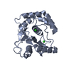
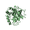
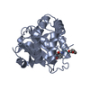
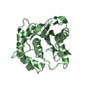
- PDB-4jkj: Crystal Structure of the S18Y Variant of Ubiquitin Carboxy-termin... -
+ Open data

- Basic information
| Entry | Database: PDB / ID: 4jkj | ||||||
|---|---|---|---|---|---|---|---|
| Title | Crystal Structure of the S18Y Variant of Ubiquitin Carboxy-terminal Hydrolase L1 | ||||||
 Components Components | Ubiquitin carboxyl-terminal hydrolase isozyme L1 | ||||||
 Keywords Keywords | HYDROLASE / Ubiquitin Hydrolase | ||||||
| Function / homology |  Function and homology information Function and homology informationmale germ cell proliferation / axon target recognition / alpha-2A adrenergic receptor binding / muscle cell development / signaling receptor inhibitor activity / adult walking behavior / neuromuscular process / axonal transport of mitochondrion / eating behavior / neuron projection terminus ...male germ cell proliferation / axon target recognition / alpha-2A adrenergic receptor binding / muscle cell development / signaling receptor inhibitor activity / adult walking behavior / neuromuscular process / axonal transport of mitochondrion / eating behavior / neuron projection terminus / regulation of macroautophagy / protein deubiquitination / negative regulation of MAPK cascade / axon cytoplasm / response to ischemia / positive regulation of glycolytic process / protein catabolic process / ubiquitin binding / cellular response to xenobiotic stimulus / UCH proteinases / ribosome binding / omega peptidase activity / proteasome-mediated ubiquitin-dependent protein catabolic process / cysteine-type deubiquitinase activity / ubiquitinyl hydrolase 1 / cysteine-type endopeptidase activity / neuronal cell body / ubiquitin protein ligase binding / endoplasmic reticulum membrane / nucleoplasm / cytosol / cytoplasm Similarity search - Function | ||||||
| Biological species |  Homo sapiens (human) Homo sapiens (human) | ||||||
| Method |  X-RAY DIFFRACTION / SYNCHROTRON / MOLECULAR REPLACEMENT / Resolution: 2.151 Å X-RAY DIFFRACTION / SYNCHROTRON / MOLECULAR REPLACEMENT / Resolution: 2.151 Å | ||||||
 Authors Authors | Davies, C.W. / Ringe, D. / Petsko, G.A. / Das, C. | ||||||
 Citation Citation | Journal: To be Published Title: Crystal Structure of the S18Y Variant of Ubiquitin Carboxy-terminal Hydrolase L1 Authors: Davies, C.W. / Ringe, D. / Petsko, G.A. / Das, C. | ||||||
| History |
|
- Structure visualization
| Structure viewer | Molecule: MolmilJmol/JSmol |
|---|
- Downloads & links
-Download
| PDBx/mmCIF format | 4jkj.cif.gz | 189.2 KB | Display | PDBx/mmCIF format |
|---|---|---|---|---|
| PDB format | pdb4jkj.ent.gz | 152.4 KB | Display | PDB format |
| PDBx/mmJSON format | 4jkj.json.gz | Tree view | PDBx/mmJSON format | |
| Others |  Other downloads Other downloads |
-Validation report
| Arichive directory | https://data.pdbj.org/pub/pdb/validation_reports/jk/4jkjftp://data.pdbj.org/pub/pdb/validation_reports/jk/4jkj | HTTPS FTP |
|---|
-Related structure data
| Related structure data |  2etlS S: Starting model for refinement |
|---|---|
| Similar structure data |


-Links
 PDBj
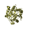
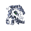
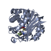
PDBj- Assembly
| Deposited unit | 
| ||||||||
|---|---|---|---|---|---|---|---|---|---|
| 1 | 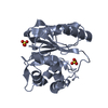
| ||||||||
| 2 | 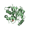
| ||||||||
| 3 | 
| ||||||||
| 4 | 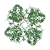
| ||||||||
| Unit cell |
|
-Components
| #1: Protein | Mass: 25342.895 Da / Num. of mol.: 2 / Mutation: S18Y Source method: isolated from a genetically manipulated source Source: (gene. exp.) Homo sapiens (human) / Gene: UCHL1 / Plasmid: pGEX-6p1 / Production host:  #2: Chemical |   Mass: 96.063 Da / Num. of mol.: 3 / Source method: obtained synthetically / Formula: SO4 Mass: 96.063 Da / Num. of mol.: 3 / Source method: obtained synthetically / Formula: SO4#3: Water | ChemComp-HOH / |  Mass: 18.015 Da / Num. of mol.: 95 / Source method: isolated from a natural source / Formula: H2O Mass: 18.015 Da / Num. of mol.: 95 / Source method: isolated from a natural source / Formula: H2O |
|---|
-Experimental details
-Experiment
| Experiment | Method: X-RAY DIFFRACTION / Number of used crystals: 1 |
|---|
- Sample preparation
| Crystal | Density Matthews: 2.36 Å3/Da / Density % sol: 47.9 % |
|---|---|
| Crystal grow | Temperature: 298 K / Method: vapor diffusion, hanging drop / pH: 7 Details: 2.4M Ammonium sulfate, 0.1M HEPES, pH 7.0, VAPOR DIFFUSION, HANGING DROP, temperature 298K |
-Data collection
| Diffraction | Mean temperature: 100 K | |||||||||||||||||||||||||||||||||||||||||||||||||
|---|---|---|---|---|---|---|---|---|---|---|---|---|---|---|---|---|---|---|---|---|---|---|---|---|---|---|---|---|---|---|---|---|---|---|---|---|---|---|---|---|---|---|---|---|---|---|---|---|---|---|
| Diffraction source | Source: SYNCHROTRON / Site: APS  / Beamline: 23-ID-D / Wavelength: 0.97 Å / Beamline: 23-ID-D / Wavelength: 0.97 Å | |||||||||||||||||||||||||||||||||||||||||||||||||
| Detector | Type: MARMOSAIC 300 mm CCD / Detector: CCD / Date: Dec 9, 2006 / Details: mirrors | |||||||||||||||||||||||||||||||||||||||||||||||||
| Radiation | Monochromator: Si 111 Channel / Protocol: SINGLE WAVELENGTH / Monochromatic (M) / Laue (L): M / Scattering type: x-ray | |||||||||||||||||||||||||||||||||||||||||||||||||
| Radiation wavelength | Wavelength: 0.97 Å / Relative weight: 1 | |||||||||||||||||||||||||||||||||||||||||||||||||
| Reflection | Resolution: 2.15→50 Å / Num. all: 26943 / Num. obs: 26890 / % possible obs: 99.9 % / Observed criterion σ(F): 4 / Observed criterion σ(I): 4 / Redundancy: 12.5 % / Rsym value: 0.064 / Net I/σ(I): 41.4 | |||||||||||||||||||||||||||||||||||||||||||||||||
| Reflection shell |
|
- Processing
| Software |
| |||||||||||||||||||||||||||||||||||||||||||||||||||||||||||||||||||||||||||||
|---|---|---|---|---|---|---|---|---|---|---|---|---|---|---|---|---|---|---|---|---|---|---|---|---|---|---|---|---|---|---|---|---|---|---|---|---|---|---|---|---|---|---|---|---|---|---|---|---|---|---|---|---|---|---|---|---|---|---|---|---|---|---|---|---|---|---|---|---|---|---|---|---|---|---|---|---|---|---|
| Refinement | Method to determine structure: MOLECULAR REPLACEMENT Starting model: PDB ENTRY 2ETL Resolution: 2.151→33.208 Å / SU ML: 0.24 / Cross valid method: THROUGHOUT / σ(F): 1.34 / Phase error: 23.51 / Stereochemistry target values: ML
| |||||||||||||||||||||||||||||||||||||||||||||||||||||||||||||||||||||||||||||
| Solvent computation | Shrinkage radii: 0.86 Å / VDW probe radii: 1.1 Å / Solvent model: FLAT BULK SOLVENT MODEL / Bsol: 54.247 Å2 / ksol: 0.394 e/Å3 | |||||||||||||||||||||||||||||||||||||||||||||||||||||||||||||||||||||||||||||
| Displacement parameters |
| |||||||||||||||||||||||||||||||||||||||||||||||||||||||||||||||||||||||||||||
| Refinement step | Cycle: LAST / Resolution: 2.151→33.208 Å
| |||||||||||||||||||||||||||||||||||||||||||||||||||||||||||||||||||||||||||||
| Refine LS restraints |
| |||||||||||||||||||||||||||||||||||||||||||||||||||||||||||||||||||||||||||||
| LS refinement shell |
| |||||||||||||||||||||||||||||||||||||||||||||||||||||||||||||||||||||||||||||
| Refinement TLS params. | Method: refined / Refine-ID: X-RAY DIFFRACTION
| |||||||||||||||||||||||||||||||||||||||||||||||||||||||||||||||||||||||||||||
| Refinement TLS group |
|
