 Movie
Movie Controller

[English] 日本語
 Yorodumi
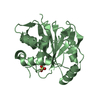
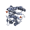
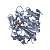
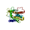
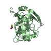
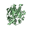
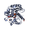
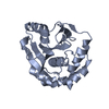
- PDB-2etl: Crystal Structure of Ubiquitin Carboxy-terminal Hydrolase L1 (UCH-L1) -
+ Open data

- Basic information
| Entry | Database: PDB / ID: 2etl | ||||||
|---|---|---|---|---|---|---|---|
| Title | Crystal Structure of Ubiquitin Carboxy-terminal Hydrolase L1 (UCH-L1) | ||||||
 Components Components | Ubiquitin carboxyl-terminal hydrolase isozyme L1 | ||||||
 Keywords Keywords | HYDROLASE / LIGASE / Deubiquitinating Thiol Hydrolase | ||||||
| Function / homology |  Function and homology information Function and homology informationaxon target recognition / male germ cell proliferation / alpha-2A adrenergic receptor binding / signaling receptor inhibitor activity / muscle cell development / adult walking behavior / neuromuscular process / axonal transport of mitochondrion / eating behavior / neuron projection terminus ...axon target recognition / male germ cell proliferation / alpha-2A adrenergic receptor binding / signaling receptor inhibitor activity / muscle cell development / adult walking behavior / neuromuscular process / axonal transport of mitochondrion / eating behavior / neuron projection terminus / regulation of macroautophagy / protein deubiquitination / negative regulation of MAPK cascade / axon cytoplasm / response to ischemia / positive regulation of glycolytic process / protein catabolic process / ubiquitin binding / cellular response to xenobiotic stimulus / UCH proteinases / ribosome binding / omega peptidase activity / proteasome-mediated ubiquitin-dependent protein catabolic process / cysteine-type deubiquitinase activity / ubiquitinyl hydrolase 1 / cysteine-type endopeptidase activity / neuronal cell body / ubiquitin protein ligase binding / endoplasmic reticulum membrane / nucleoplasm / cytoplasm / cytosol Similarity search - Function | ||||||
| Biological species |  Homo sapiens (human) Homo sapiens (human) | ||||||
| Method |  X-RAY DIFFRACTION / SYNCHROTRON / MOLECULAR REPLACEMENT / Resolution: 2.4 Å X-RAY DIFFRACTION / SYNCHROTRON / MOLECULAR REPLACEMENT / Resolution: 2.4 Å | ||||||
 Authors Authors | Das, C. / Hoang, Q.Q. / Kreinbring, C.A. / Luchansky, S.J. / Meray, R.K. / Ray, S.S. / Lansbury, P.T. / Ringe, D. / Petsko, G.A. | ||||||
 Citation Citation | Journal: Proc.Natl.Acad.Sci.USA / Year: 2006 Title: Structural basis for conformational plasticity of the Parkinson's disease-associated ubiquitin hydrolase UCH-L1. Authors: Das, C. / Hoang, Q.Q. / Kreinbring, C.A. / Luchansky, S.J. / Meray, R.K. / Ray, S.S. / Lansbury, P.T. / Ringe, D. / Petsko, G.A. | ||||||
| History |
|
- Structure visualization
| Structure viewer | Molecule: MolmilJmol/JSmol |
|---|
- Downloads & links
-Download
| PDBx/mmCIF format | 2etl.cif.gz | 99.3 KB | Display | PDBx/mmCIF format |
|---|---|---|---|---|
| PDB format | pdb2etl.ent.gz | 76.5 KB | Display | PDB format |
| PDBx/mmJSON format | 2etl.json.gz | Tree view | PDBx/mmJSON format | |
| Others |  Other downloads Other downloads |
-Validation report
| Arichive directory | https://data.pdbj.org/pub/pdb/validation_reports/et/2etlftp://data.pdbj.org/pub/pdb/validation_reports/et/2etl | HTTPS FTP |
|---|
-Related structure data
| Similar structure data |
|---|


-Links
 PDBj
PDBj- Assembly
| Deposited unit | 
| ||||||||
|---|---|---|---|---|---|---|---|---|---|
| 1 | 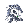
| ||||||||
| 2 | 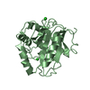
| ||||||||
| 3 | 
| ||||||||
| 4 | 
| ||||||||
| Unit cell |
| ||||||||
| Details | The biological assembly is a monomer, a subunit of the assymetric unit which contains two copies of the protein molecule. |
-Components
| #1: Protein | Mass: 25266.799 Da / Num. of mol.: 2 Source method: isolated from a genetically manipulated source Source: (gene. exp.) Homo sapiens (human) / Gene: UCHL1 / Plasmid: pGEX-6P1 / Production host:  References: UniProt: P09936, ubiquitinyl hydrolase 1, Ligases #2: Chemical | ChemComp-CL /   Mass: 35.453 Da / Num. of mol.: 4 / Source method: obtained synthetically / Formula: Cl Mass: 35.453 Da / Num. of mol.: 4 / Source method: obtained synthetically / Formula: Cl#3: Water | ChemComp-HOH / |  Mass: 18.015 Da / Num. of mol.: 107 / Source method: isolated from a natural source / Formula: H2O Mass: 18.015 Da / Num. of mol.: 107 / Source method: isolated from a natural source / Formula: H2O |
|---|
-Experimental details
-Experiment
| Experiment | Method: X-RAY DIFFRACTION / Number of used crystals: 1 |
|---|
- Sample preparation
| Crystal | Density Matthews: 2.38 Å3/Da / Density % sol: 48.37 % |
|---|---|
| Crystal grow | Temperature: 293 K / Method: vapor diffusion, hanging drop / pH: 7 Details: 2.4 M Ammonium Sulfate, 0.1 M HEPES, pH 7.0, VAPOR DIFFUSION, HANGING DROP, temperature 293.0K |
-Data collection
| Diffraction | Mean temperature: 298 K |
|---|---|
| Diffraction source | Source: SYNCHROTRON / Site: SSRL  / Beamline: BL9-1 / Wavelength: 0.98397 Å / Beamline: BL9-1 / Wavelength: 0.98397 Å |
| Detector | Type: ADSC QUANTUM 315 / Detector: CCD / Date: Apr 29, 2005 / Details: Flat mirror (vertical focusing) |
| Radiation | Monochromator: Si(311) / Protocol: SINGLE WAVELENGTH / Monochromatic (M) / Laue (L): M / Scattering type: x-ray |
| Radiation wavelength | Wavelength: 0.98397 Å / Relative weight: 1 |
| Reflection | Resolution: 2.4→41.9 Å / Num. all: 34753 / Num. obs: 34753 / % possible obs: 100 % / Observed criterion σ(F): 3 / Observed criterion σ(I): 3 / Redundancy: 14.1 % / Rmerge(I) obs: 0.088 / Χ2: 0.997 |
| Reflection shell | Resolution: 2.4→2.49 Å / % possible obs: 99.7 % / Redundancy: 14.3 % / Rmerge(I) obs: 0.773 / Num. measured obs: 1925 / Χ2: 1.17 / % possible all: 99.7 |
-Phasing
| Phasing MR | Rfactor: 0.554 / Cor.coef. Fo:Fc: 0.284
|
|---|
- Processing
| Software |
| ||||||||||||||||||||||||||||
|---|---|---|---|---|---|---|---|---|---|---|---|---|---|---|---|---|---|---|---|---|---|---|---|---|---|---|---|---|---|
| Refinement | Method to determine structure: MOLECULAR REPLACEMENT Starting model: Homology model based on UCH-L3 Resolution: 2.4→41.9 Å / Cross valid method: THROUGHOUT / σ(F): 0 / σ(I): 0 / Stereochemistry target values: Engh & Huber
| ||||||||||||||||||||||||||||
| Solvent computation | Bsol: 47.87 Å2 | ||||||||||||||||||||||||||||
| Displacement parameters | Biso mean: 49.283 Å2
| ||||||||||||||||||||||||||||
| Refinement step | Cycle: LAST / Resolution: 2.4→41.9 Å
| ||||||||||||||||||||||||||||
| Refine LS restraints |
| ||||||||||||||||||||||||||||
| Xplor file |
|
