 Movie
Movie Controller

[English] 日本語
 Yorodumi
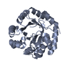
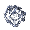
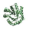
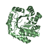
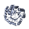
- PDB-2dzx: Structure of mutant tryptophan synthase alpha-subunit (E131-132A)... -
+ Open data

- Basic information
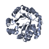
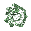
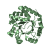
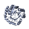
| Entry | Database: PDB / ID: 2dzx | ||||||
|---|---|---|---|---|---|---|---|
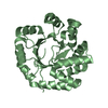
| Title | Structure of mutant tryptophan synthase alpha-subunit (E131-132A) from a hyperthermophile, Pyrococcus furiosus | ||||||
 Components Components | Tryptophan synthase alpha chain | ||||||
 Keywords Keywords | LYASE / tryptophan synthase alpha-subunit / hyperthermophile / pyrococcus furiosus / x-ray analysis / stability / calorimetry / Structural Genomics / NPPSFA / National Project on Protein Structural and Functional Analyses / RIKEN Structural Genomics/Proteomics Initiative / RSGI | ||||||
| Function / homology |  Function and homology information Function and homology information | ||||||
| Biological species |   Pyrococcus furiosus (archaea) Pyrococcus furiosus (archaea) | ||||||
| Method |  X-RAY DIFFRACTION / SYNCHROTRON / FOURIER SYNTHESIS / Resolution: 2.4 Å X-RAY DIFFRACTION / SYNCHROTRON / FOURIER SYNTHESIS / Resolution: 2.4 Å | ||||||
 Authors Authors | Ogasahara, K. / Yamagata, Y. / Yutani, K. / RIKEN Structural Genomics/Proteomics Initiative (RSGI) | ||||||
 Citation Citation | Journal: To be Published Title: Structures of mutant tryptophan synthase alpha-subunits from a hyperthermophile, Pyrococcus furiosus Authors: Ogasahara, K. / Yamagata, Y. / Yutani, K. #1: Journal: J.Biol.Chem. / Year: 2001Title: Entropic stabilization of the tryptophan synthase alpha-subunit from a hyperthermophile, Pyrococcus furiosus. X-ray analysis and calorimetry Authors: Yamagata, Y. / Ogasahara, K. / Hioki, Y. / Lee, S.J. / Nakagawa, A. / Nakamura, H. / Ishida, M. / Kuramitsu, S. / Yutani, K. | ||||||
| History |
|
- Structure visualization
| Structure viewer | Molecule: MolmilJmol/JSmol |
|---|
- Downloads & links
-Download
| PDBx/mmCIF format | 2dzx.cif.gz | 114.8 KB | Display | PDBx/mmCIF format |
|---|---|---|---|---|
| PDB format | pdb2dzx.ent.gz | 89.9 KB | Display | PDB format |
| PDBx/mmJSON format | 2dzx.json.gz | Tree view | PDBx/mmJSON format | |
| Others |  Other downloads Other downloads |
-Validation report
| Arichive directory | https://data.pdbj.org/pub/pdb/validation_reports/dz/2dzxftp://data.pdbj.org/pub/pdb/validation_reports/dz/2dzx | HTTPS FTP |
|---|
-Related structure data
| Related structure data | 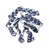 2dzpC 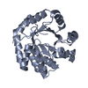 2dzsC  2dztC 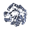 2dzuC  2dzvC  2dzwC  2e09C  1geqS S: Starting model for refinement C: citing same article ( |
|---|---|
| Similar structure data | |
| Other databases |


-Links
 PDBj
PDBj
- Assembly
| Deposited unit | 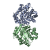
| ||||||||
|---|---|---|---|---|---|---|---|---|---|
| 1 | 
| ||||||||
| 2 | 
| ||||||||
| Unit cell |
|
-Components
| #1: Protein | Mass: 27419.773 Da / Num. of mol.: 2 / Mutation: E131A,E132A Source method: isolated from a genetically manipulated source Source: (gene. exp.) Pyrococcus furiosus (archaea) / Gene: trpA / Plasmid: ps1974 / Production host:  #2: Water | ChemComp-HOH / |  Mass: 18.015 Da / Num. of mol.: 474 / Source method: isolated from a natural source / Formula: H2O Mass: 18.015 Da / Num. of mol.: 474 / Source method: isolated from a natural source / Formula: H2O |
|---|
-Experimental details
-Experiment
| Experiment | Method: X-RAY DIFFRACTION / Number of used crystals: 1 |
|---|
- Sample preparation
| Crystal | Density Matthews: 2.17 Å3/Da / Density % sol: 43.33 % |
|---|---|
| Crystal grow | Temperature: 283 K / Method: vapor diffusion, hanging drop / pH: 6.5 Details: 0.1M MES-NAOH, 12% PEG 20000, pH 6.5, VAPOR DIFFUSION, HANGING DROP, temperature 283K |
-Data collection
| Diffraction | Mean temperature: 100 K |
|---|---|
| Diffraction source | Source: SYNCHROTRON / Site: SPring-8  / Beamline: BL41XU / Wavelength: 1 Å / Beamline: BL41XU / Wavelength: 1 Å |
| Detector | Type: MAR CCD 165 mm / Detector: CCD / Date: May 16, 2001 / Details: mirror |
| Radiation | Monochromator: Si / Protocol: SINGLE WAVELENGTH / Monochromatic (M) / Laue (L): M / Scattering type: x-ray |
| Radiation wavelength | Wavelength: 1 Å / Relative weight: 1 |
| Reflection | Resolution: 2.19→99 Å / Num. obs: 23394 / % possible obs: 94 % / Observed criterion σ(F): 0 / Observed criterion σ(I): 0 / Redundancy: 6.56 % / Biso Wilson estimate: 27.8 Å2 / Rmerge(I) obs: 0.054 / Net I/σ(I): 42.1 |
| Reflection shell | Resolution: 2.19→2.27 Å / Rmerge(I) obs: 0.171 / Mean I/σ(I) obs: 4 / Num. unique all: 1846 / % possible all: 75.7 |
- Processing
| Software |
| ||||||||||||||||||||||||||||||||||||||||||||||||||||||||||||||||||||||||||||||||
|---|---|---|---|---|---|---|---|---|---|---|---|---|---|---|---|---|---|---|---|---|---|---|---|---|---|---|---|---|---|---|---|---|---|---|---|---|---|---|---|---|---|---|---|---|---|---|---|---|---|---|---|---|---|---|---|---|---|---|---|---|---|---|---|---|---|---|---|---|---|---|---|---|---|---|---|---|---|---|---|---|---|
| Refinement | Method to determine structure: FOURIER SYNTHESIS Starting model: 1GEQ Resolution: 2.4→19.73 Å / Rfactor Rfree error: 0.009 / Data cutoff high absF: 1427736.69 / Data cutoff low absF: 0 / Isotropic thermal model: RESTRAINED / Cross valid method: THROUGHOUT / σ(F): 0 / σ(I): 0
| ||||||||||||||||||||||||||||||||||||||||||||||||||||||||||||||||||||||||||||||||
| Solvent computation | Solvent model: FLAT MODEL / Bsol: 45.0651 Å2 / ksol: 0.335521 e/Å3 | ||||||||||||||||||||||||||||||||||||||||||||||||||||||||||||||||||||||||||||||||
| Displacement parameters | Biso mean: 22.1 Å2
| ||||||||||||||||||||||||||||||||||||||||||||||||||||||||||||||||||||||||||||||||
| Refine analyze |
| ||||||||||||||||||||||||||||||||||||||||||||||||||||||||||||||||||||||||||||||||
| Refinement step | Cycle: LAST / Resolution: 2.4→19.73 Å
| ||||||||||||||||||||||||||||||||||||||||||||||||||||||||||||||||||||||||||||||||
| Refine LS restraints |
| ||||||||||||||||||||||||||||||||||||||||||||||||||||||||||||||||||||||||||||||||
| LS refinement shell | Resolution: 2.4→2.55 Å / Rfactor Rfree error: 0.024 / Total num. of bins used: 6
| ||||||||||||||||||||||||||||||||||||||||||||||||||||||||||||||||||||||||||||||||
| Xplor file |
|
