 Movie
Movie Controller

[English] 日本語
 Yorodumi
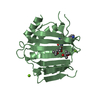
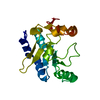
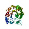
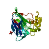
- PDB-1wxj: Crystal Structure Of Tryptophan Synthase A-Subunit with Indole-3-... -
+ Open data

- Basic information
| Entry | Database: PDB / ID: 1wxj | ||||||
|---|---|---|---|---|---|---|---|
| Title | Crystal Structure Of Tryptophan Synthase A-Subunit with Indole-3-propanol phosphate From Thermus Thermophilus Hb8 | ||||||
 Components Components | tryptophan synthase alpha chain | ||||||
 Keywords Keywords | LYASE / structural genomics / Thermus thermophilus HB8 / RIKEN Structural Genomics/Proteomics Initiative / RSGI | ||||||
| Function / homology |  Function and homology information Function and homology information | ||||||
| Biological species |   Thermus thermophilus (bacteria) Thermus thermophilus (bacteria) | ||||||
| Method |  X-RAY DIFFRACTION / SYNCHROTRON / MOLECULAR REPLACEMENT / Resolution: 1.7 Å X-RAY DIFFRACTION / SYNCHROTRON / MOLECULAR REPLACEMENT / Resolution: 1.7 Å | ||||||
 Authors Authors | Asada, Y. / Kunishima, N. / RIKEN Structural Genomics/Proteomics Initiative (RSGI) | ||||||
 Citation Citation | Journal: To be Published Title: Crystal Structure Of Tryptophan Synthase A-Subunit with Indole-3-propanol phosphate From Thermus Thermophilus Hb8 Authors: Asada, Y. / Kunishima, N. | ||||||
| History |
|
- Structure visualization
| Structure viewer | Molecule: MolmilJmol/JSmol |
|---|
- Downloads & links
-Download
| PDBx/mmCIF format | 1wxj.cif.gz | 66.9 KB | Display | PDBx/mmCIF format |
|---|---|---|---|---|
| PDB format | pdb1wxj.ent.gz | 47.2 KB | Display | PDB format |
| PDBx/mmJSON format | 1wxj.json.gz | Tree view | PDBx/mmJSON format | |
| Others |  Other downloads Other downloads |
-Validation report
| Arichive directory | https://data.pdbj.org/pub/pdb/validation_reports/wx/1wxjftp://data.pdbj.org/pub/pdb/validation_reports/wx/1wxj | HTTPS FTP |
|---|
-Related structure data
| Related structure data |  1ujpS S: Starting model for refinement |
|---|---|
| Similar structure data | |
| Other databases |


-Links
 PDBj
PDBj
- Assembly
| Deposited unit | 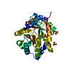
| ||||||||
|---|---|---|---|---|---|---|---|---|---|
| 1 |
| ||||||||
| Unit cell |
| ||||||||
| Details | The biological assembly is monomer |
-Components
| #1: Protein | Mass: 28977.525 Da / Num. of mol.: 1 Source method: isolated from a genetically manipulated source Source: (gene. exp.) Thermus thermophilus (bacteria) / Strain: HB8 / Plasmid: pET11a / Species (production host): Escherichia coli / Production host: | ||||
|---|---|---|---|---|---|
| #2: Chemical | ChemComp-SO4 /   Mass: 96.063 Da / Num. of mol.: 4 / Source method: obtained synthetically / Formula: SO4 Mass: 96.063 Da / Num. of mol.: 4 / Source method: obtained synthetically / Formula: SO4#3: Chemical | ChemComp-IPL / | 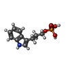  Mass: 255.207 Da / Num. of mol.: 1 / Source method: obtained synthetically / Formula: C11H14NO4P Mass: 255.207 Da / Num. of mol.: 1 / Source method: obtained synthetically / Formula: C11H14NO4P#4: Water | ChemComp-HOH / |  Mass: 18.015 Da / Num. of mol.: 278 / Source method: isolated from a natural source / Formula: H2O Mass: 18.015 Da / Num. of mol.: 278 / Source method: isolated from a natural source / Formula: H2O |
-Experimental details
-Experiment
| Experiment | Method: X-RAY DIFFRACTION / Number of used crystals: 1 |
|---|
- Sample preparation
| Crystal | Density Matthews: 2.16 Å3/Da / Density % sol: 43 % |
|---|---|
| Crystal grow | Temperature: 291 K / Method: microbatch / pH: 8.5 Details: INDOLE-3-PROPANOL PHOSPHATE, Tris-HCl, Ammonium Sulfate, pH 8.5, MICROBATCH, temperature 291K |
-Data collection
| Diffraction | Mean temperature: 100 K |
|---|---|
| Diffraction source | Source: SYNCHROTRON / Site: SPring-8  / Beamline: BL26B1 / Wavelength: 0.9 Å / Beamline: BL26B1 / Wavelength: 0.9 Å |
| Detector | Type: RIGAKU JUPITER 210 / Detector: CCD / Date: Apr 23, 2004 / Details: mirrors |
| Radiation | Monochromator: Bending Magnet / Protocol: SINGLE WAVELENGTH / Monochromatic (M) / Laue (L): M / Scattering type: x-ray |
| Radiation wavelength | Wavelength: 0.9 Å / Relative weight: 1 |
| Reflection | Resolution: 1.7→50 Å / Num. all: 26921 / Num. obs: 26921 / % possible obs: 100 % / Observed criterion σ(F): 0 / Observed criterion σ(I): 0 / Redundancy: 3.8 % / Biso Wilson estimate: 18.927 Å2 / Rmerge(I) obs: 0.044 / Rsym value: 0.038 / Net I/σ(I): 15.1 |
| Reflection shell | Resolution: 1.7→1.76 Å / Redundancy: 3.8 % / Rmerge(I) obs: 0.297 / Mean I/σ(I) obs: 4.01 / Num. unique all: 2664 / Rsym value: 0.253 / % possible all: 100 |
- Processing
| Software |
| |||||||||||||||||||||||||
|---|---|---|---|---|---|---|---|---|---|---|---|---|---|---|---|---|---|---|---|---|---|---|---|---|---|---|
| Refinement | Method to determine structure: MOLECULAR REPLACEMENT Starting model: 1UJP Resolution: 1.7→27.08 Å / Isotropic thermal model: RESTRAINED / Cross valid method: THROUGHOUT / σ(F): 0 / Stereochemistry target values: Engh & Huber
| |||||||||||||||||||||||||
| Displacement parameters | Biso mean: 23.3 Å2
| |||||||||||||||||||||||||
| Refine analyze |
| |||||||||||||||||||||||||
| Refinement step | Cycle: LAST / Resolution: 1.7→27.08 Å
| |||||||||||||||||||||||||
| Refine LS restraints |
| |||||||||||||||||||||||||
| LS refinement shell | Resolution: 1.7→1.78 Å / Rfactor Rfree error: 0.022
|
