 Movie
Movie Controller

[English] 日本語
 Yorodumi
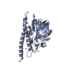
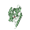
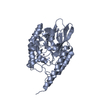
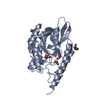
- PDB-1ujp: Crystal Structure of Tryptophan Synthase A-Subunit From Thermus t... -
+ Open data

- Basic information
| Entry | Database: PDB / ID: 1ujp | ||||||
|---|---|---|---|---|---|---|---|
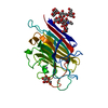
| Title | Crystal Structure of Tryptophan Synthase A-Subunit From Thermus thermophilus HB8 | ||||||
 Components Components | Tryptophan synthase alpha chain | ||||||
 Keywords Keywords | LYASE / Tryptophan Synthase / Tryptophan / RIKEN Structural Genomics/Proteomics Initiative / RSGI / Structural Genomics | ||||||
| Function / homology |  Function and homology information Function and homology information | ||||||
| Biological species |   Thermus thermophilus (bacteria) Thermus thermophilus (bacteria) | ||||||
| Method |  X-RAY DIFFRACTION / SYNCHROTRON / MOLECULAR REPLACEMENT / Resolution: 1.34 Å X-RAY DIFFRACTION / SYNCHROTRON / MOLECULAR REPLACEMENT / Resolution: 1.34 Å | ||||||
 Authors Authors | Asada, Y. / Yokoyama, S. / Kuramitsu, S. / Miyano, M. / Kunishima, N. / RIKEN Structural Genomics/Proteomics Initiative (RSGI) | ||||||
 Citation Citation | Journal: J.Biochem.(Tokyo) / Year: 2005 Title: Stabilization mechanism of the tryptophan synthase alpha-subunit from Thermus thermophilus HB8: X-ray crystallographic analysis and calorimetry. Authors: Asada, Y. / Sawano, M. / Ogasahara, K. / Nakamura, J. / Ota, M. / Kuroishi, C. / Sugahara, M. / Yutani, K. / Kunishima, N. | ||||||
| History |
|
- Structure visualization
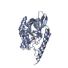
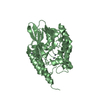
| Structure viewer | Molecule: MolmilJmol/JSmol |
|---|
- Downloads & links
-Download
| PDBx/mmCIF format | 1ujp.cif.gz | 66.8 KB | Display | PDBx/mmCIF format |
|---|---|---|---|---|
| PDB format | pdb1ujp.ent.gz | 47.6 KB | Display | PDB format |
| PDBx/mmJSON format | 1ujp.json.gz | Tree view | PDBx/mmJSON format | |
| Others |  Other downloads Other downloads |
-Validation report
| Arichive directory | https://data.pdbj.org/pub/pdb/validation_reports/uj/1ujpftp://data.pdbj.org/pub/pdb/validation_reports/uj/1ujp | HTTPS FTP |
|---|
-Related structure data
| Related structure data |  1geqS S: Starting model for refinement |
|---|---|
| Similar structure data | |
| Other databases |

-Links
 PDBj
PDBj
- Assembly
| Deposited unit | 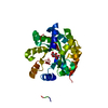
| ||||||||
|---|---|---|---|---|---|---|---|---|---|
| 1 |
| ||||||||
| Unit cell |
|
-Components
| #1: Protein | Mass: 28977.525 Da / Num. of mol.: 1 Source method: isolated from a genetically manipulated source Source: (gene. exp.) Thermus thermophilus (bacteria) / Strain: HB8 / Plasmid: pET11a / Production host: |
|---|---|
| #2: Chemical | ChemComp-CIT /   Mass: 192.124 Da / Num. of mol.: 1 / Source method: obtained synthetically / Formula: C6H8O7 Mass: 192.124 Da / Num. of mol.: 1 / Source method: obtained synthetically / Formula: C6H8O7 |
| #3: Water | ChemComp-HOH /  Mass: 18.015 Da / Num. of mol.: 303 / Source method: isolated from a natural source / Formula: H2O Mass: 18.015 Da / Num. of mol.: 303 / Source method: isolated from a natural source / Formula: H2O |
-Experimental details
-Experiment
| Experiment | Method: X-RAY DIFFRACTION / Number of used crystals: 1 |
|---|
- Sample preparation
| Crystal | Density Matthews: 2.07 Å3/Da / Density % sol: 40.18 % | ||||||||||||||||||||||||||||||||||||||||||||||||||||||||
|---|---|---|---|---|---|---|---|---|---|---|---|---|---|---|---|---|---|---|---|---|---|---|---|---|---|---|---|---|---|---|---|---|---|---|---|---|---|---|---|---|---|---|---|---|---|---|---|---|---|---|---|---|---|---|---|---|---|
| Crystal grow | Temperature: 291 K / Method: microbatch / pH: 5.6 Details: Ammonium Acetate, tri-Sodium Citrate dihydrate, PEG 4000, Glycerol anhydrous, pH 5.6, MICROBATCH, temperature 291K | ||||||||||||||||||||||||||||||||||||||||||||||||||||||||
| Crystal grow | *PLUS Temperature: 291 K / pH: 8 / Method: batch method | ||||||||||||||||||||||||||||||||||||||||||||||||||||||||
| Components of the solutions | *PLUS
|
-Data collection
| Diffraction | Mean temperature: 100 K |
|---|---|
| Diffraction source | Source: SYNCHROTRON / Site: SPring-8  / Beamline: BL26B1 / Wavelength: 0.8 Å / Beamline: BL26B1 / Wavelength: 0.8 Å |
| Detector | Type: RIGAKU RAXIS V / Detector: IMAGE PLATE / Date: Jun 5, 2003 / Details: mirrors |
| Radiation | Monochromator: Bending Magnet / Protocol: SINGLE WAVELENGTH / Monochromatic (M) / Laue (L): M / Scattering type: x-ray |
| Radiation wavelength | Wavelength: 0.8 Å / Relative weight: 1 |
| Reflection | Resolution: 1.34→30 Å / Num. all: 55454 / Num. obs: 55454 / % possible obs: 99.6 % / Observed criterion σ(F): 0 / Observed criterion σ(I): 0 / Redundancy: 3.6 % / Biso Wilson estimate: 13.6 Å2 / Rmerge(I) obs: 0.045 / Rsym value: 0.042 / Net I/σ(I): 14.1 |
| Reflection shell | Resolution: 1.34→1.39 Å / Redundancy: 3.7 % / Rmerge(I) obs: 0.356 / Mean I/σ(I) obs: 3.6 / Num. unique all: 5551 / Rsym value: 0.304 / % possible all: 100 |
| Reflection | *PLUS Num. measured all: 201760 |
| Reflection shell | *PLUS % possible obs: 100 % |
- Processing
| Software |
| |||||||||||||||||||||||||
|---|---|---|---|---|---|---|---|---|---|---|---|---|---|---|---|---|---|---|---|---|---|---|---|---|---|---|
| Refinement | Method to determine structure: MOLECULAR REPLACEMENT Starting model: PDB ENTRY 1GEQ Resolution: 1.34→27.3 Å / Isotropic thermal model: Anisotropic / Cross valid method: THROUGHOUT / σ(F): 0 / Stereochemistry target values: Engh & Huber
| |||||||||||||||||||||||||
| Displacement parameters | Biso mean: 17.3 Å2
| |||||||||||||||||||||||||
| Refine analyze |
| |||||||||||||||||||||||||
| Refinement step | Cycle: LAST / Resolution: 1.34→27.3 Å
| |||||||||||||||||||||||||
| Refine LS restraints |
| |||||||||||||||||||||||||
| LS refinement shell | Resolution: 1.34→1.4 Å / Rfactor Rfree error: 0.015
| |||||||||||||||||||||||||
| Refinement | *PLUS Lowest resolution: 30 Å / % reflection Rfree: 5 % | |||||||||||||||||||||||||
| Solvent computation | *PLUS | |||||||||||||||||||||||||
| Displacement parameters | *PLUS | |||||||||||||||||||||||||
| LS refinement shell | *PLUS Lowest resolution: 1.39 Å |
