 Movie
Movie Controller


+ Open data

- Basic information
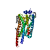
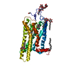
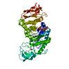
| Entry | Database: PDB / ID: 2efw | ||||||
|---|---|---|---|---|---|---|---|
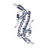
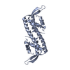
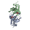
| Title | Crystal structure of the RTP:nRB complex from Bacillus subtilis | ||||||
 Components Components |
| ||||||
 Keywords Keywords | REPLICATION/DNA / protein-DNA complex / 'winged'-helix protein / DNA replication termination / replication fork arrest / REPLICATION-DNA COMPLEX | ||||||
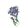
| Function / homology |  Function and homology information Function and homology information | ||||||
| Biological species |  | ||||||
| Method |  X-RAY DIFFRACTION / SYNCHROTRON / MOLECULAR REPLACEMENT / Resolution: 2.5 Å X-RAY DIFFRACTION / SYNCHROTRON / MOLECULAR REPLACEMENT / Resolution: 2.5 Å | ||||||
 Authors Authors | Vivian, J.P. / Porter, C.J. / Wilce, J.A. / Wilce, M.C.J. | ||||||
 Citation Citation | Journal: J.Mol.Biol. / Year: 2007 Title: An asymmetric structure of the Bacillus subtilis replication terminator protein in complex with DNA Authors: Vivian, J.P. / Porter, C.J. / Wilce, J.A. / Wilce, M.C.J. | ||||||
| History |
|
- Structure visualization
| Structure viewer | Molecule: MolmilJmol/JSmol |
|---|
- Downloads & links
-Download
| PDBx/mmCIF format | 2efw.cif.gz | 143.4 KB | Display | PDBx/mmCIF format |
|---|---|---|---|---|
| PDB format | pdb2efw.ent.gz | 109.6 KB | Display | PDB format |
| PDBx/mmJSON format | 2efw.json.gz | Tree view | PDBx/mmJSON format | |
| Others |  Other downloads Other downloads |
-Validation report
| Summary document | 2efw_validation.pdf.gz | 482.7 KB | Display | wwPDB validaton report |
|---|---|---|---|---|
| Full document | 2efw_full_validation.pdf.gz | 554 KB | Display | |
| Data in XML | 2efw_validation.xml.gz | 29.1 KB | Display | |
| Data in CIF | 2efw_validation.cif.gz | 39.9 KB | Display | |
| Arichive directory | https://data.pdbj.org/pub/pdb/validation_reports/ef/2efwftp://data.pdbj.org/pub/pdb/validation_reports/ef/2efw | HTTPS FTP |
-Related structure data
| Related structure data | 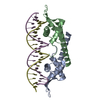 1f4kS S: Starting model for refinement |
|---|---|
| Similar structure data |


-Links
 PDBj
PDBj


- Assembly
| Deposited unit | 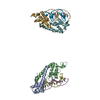
| ||||||||
|---|---|---|---|---|---|---|---|---|---|
| 1 | 
| ||||||||
| 2 | 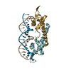
| ||||||||
| Unit cell |
|
-Components
| #1: DNA chain | Mass: 6397.159 Da / Num. of mol.: 2 / Source method: obtained synthetically / Details: the B site sequence of TerI #2: DNA chain | Mass: 6486.222 Da / Num. of mol.: 2 / Source method: obtained synthetically / Details: the B site sequence of TerI #3: Protein | Mass: 14531.099 Da / Num. of mol.: 4 / Mutation: C110S Source method: isolated from a genetically manipulated source Source: (gene. exp.) #4: Water | ChemComp-HOH / |  Mass: 18.015 Da / Num. of mol.: 24 / Source method: isolated from a natural source / Formula: H2O Mass: 18.015 Da / Num. of mol.: 24 / Source method: isolated from a natural source / Formula: H2O |
|---|
-Experimental details
-Experiment
| Experiment | Method: X-RAY DIFFRACTION / Number of used crystals: 1 |
|---|
- Sample preparation
| Crystal | Density Matthews: 2.97 Å3/Da / Density % sol: 58.54 % | ||||||||||||||||||||||||||||
|---|---|---|---|---|---|---|---|---|---|---|---|---|---|---|---|---|---|---|---|---|---|---|---|---|---|---|---|---|---|
| Crystal grow | Temperature: 295 K / Method: vapor diffusion, hanging drop / pH: 4.6 Details: 2% PEG 4000, 125mM sodium acetate, pH 4.6, VAPOR DIFFUSION, HANGING DROP, temperature 295K | ||||||||||||||||||||||||||||
| Components of the solutions |
|
-Data collection
| Diffraction | Mean temperature: 100 K |
|---|---|
| Diffraction source | Source: SYNCHROTRON / Site: APS  / Beamline: 14-BM-C / Wavelength: 0.9 Å / Beamline: 14-BM-C / Wavelength: 0.9 Å |
| Detector | Type: ADSC QUANTUM 4 / Detector: CCD / Date: Mar 15, 2003 |
| Radiation | Protocol: SINGLE WAVELENGTH / Monochromatic (M) / Laue (L): M / Scattering type: x-ray |
| Radiation wavelength | Wavelength: 0.9 Å / Relative weight: 1 |
| Reflection | Resolution: 2.5→50 Å / Num. obs: 32161 / % possible obs: 94.3 % / Observed criterion σ(F): 1 / Observed criterion σ(I): 1 / Redundancy: 4.7 % / Biso Wilson estimate: 52.4 Å2 / Rmerge(I) obs: 0.091 / Net I/σ(I): 12.7 |
| Reflection shell | Resolution: 2.5→2.56 Å / Redundancy: 2.1 % / Rmerge(I) obs: 0.341 / Mean I/σ(I) obs: 2.3 / % possible all: 84.8 |
- Processing
| Software |
| ||||||||||||||||||||||||||||||||||||||||||||||||||||||||||||||||||||||||||||||||||||||||||
|---|---|---|---|---|---|---|---|---|---|---|---|---|---|---|---|---|---|---|---|---|---|---|---|---|---|---|---|---|---|---|---|---|---|---|---|---|---|---|---|---|---|---|---|---|---|---|---|---|---|---|---|---|---|---|---|---|---|---|---|---|---|---|---|---|---|---|---|---|---|---|---|---|---|---|---|---|---|---|---|---|---|---|---|---|---|---|---|---|---|---|---|
| Refinement | Method to determine structure: MOLECULAR REPLACEMENT Starting model: PDB ENTRY 1F4K (with residues 72 to 88 and the DNA removed) Resolution: 2.5→50 Å / Cor.coef. Fo:Fc: 0.881 / Cor.coef. Fo:Fc free: 0.855 / SU B: 31.275 / SU ML: 0.321 / TLS residual ADP flag: LIKELY RESIDUAL / Cross valid method: THROUGHOUT / ESU R: 0.53 / ESU R Free: 0.349 / Stereochemistry target values: MAXIMUM LIKELIHOOD
| ||||||||||||||||||||||||||||||||||||||||||||||||||||||||||||||||||||||||||||||||||||||||||
| Solvent computation | Ion probe radii: 0.8 Å / Shrinkage radii: 0.8 Å / VDW probe radii: 1.2 Å / Solvent model: MASK | ||||||||||||||||||||||||||||||||||||||||||||||||||||||||||||||||||||||||||||||||||||||||||
| Displacement parameters | Biso mean: 11.106 Å2
| ||||||||||||||||||||||||||||||||||||||||||||||||||||||||||||||||||||||||||||||||||||||||||
| Refinement step | Cycle: LAST / Resolution: 2.5→50 Å
| ||||||||||||||||||||||||||||||||||||||||||||||||||||||||||||||||||||||||||||||||||||||||||
| Refine LS restraints |
| ||||||||||||||||||||||||||||||||||||||||||||||||||||||||||||||||||||||||||||||||||||||||||
| LS refinement shell | Resolution: 2.5→2.565 Å / Total num. of bins used: 20
| ||||||||||||||||||||||||||||||||||||||||||||||||||||||||||||||||||||||||||||||||||||||||||
| Refinement TLS params. | Method: refined / Refine-ID: X-RAY DIFFRACTION
| ||||||||||||||||||||||||||||||||||||||||||||||||||||||||||||||||||||||||||||||||||||||||||
| Refinement TLS group |
|
