 Movie
Movie Controller

[English] 日本語
 Yorodumi
- PDB-1f4k: CRYSTAL STRUCTURE OF THE REPLICATION TERMINATOR PROTEIN/B-SITE DN... -
+ Open data

- Basic information
| Entry | Database: PDB / ID: 1f4k | ||||||
|---|---|---|---|---|---|---|---|
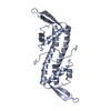
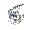
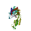
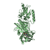
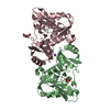
| Title | CRYSTAL STRUCTURE OF THE REPLICATION TERMINATOR PROTEIN/B-SITE DNA COMPLEX | ||||||
 Components Components |
| ||||||
 Keywords Keywords | REPLICATION/DNA / winged-helix protein-DNA complex / REPLICATION AND TERMINATION / FORK ARREST MECHANISM / REPLICATION-DNA COMPLEX | ||||||
| Function / homology |  Function and homology information Function and homology information | ||||||
| Biological species |  | ||||||
| Method |  X-RAY DIFFRACTION / SYNCHROTRON / Resolution: 2.5 Å X-RAY DIFFRACTION / SYNCHROTRON / Resolution: 2.5 Å | ||||||
 Authors Authors | Wilce, J.A. / Vivian, J.P. / Hastings, A.F. / Otting, G. / Folmer, R.H.A. / Duggin, I.G. / Wake, R.G. / Wilce, M.C.J. | ||||||
 Citation Citation | Journal: Nat.Struct.Biol. / Year: 2001 Title: Structure of the RTP-DNA complex and the mechanism of polar replication fork arrest Authors: Wilce, J.A. / Vivian, J.P. / Hastings, A.F. / Otting, G. / Folmer, R.H. / Duggin, I.G. / Wake, R.G. / Wilce, M.C. | ||||||
| History |
|
- Structure visualization
| Structure viewer | Molecule: MolmilJmol/JSmol |
|---|
- Downloads & links
-Download
| PDBx/mmCIF format | 1f4k.cif.gz | 84.1 KB | Display | PDBx/mmCIF format |
|---|---|---|---|---|
| PDB format | pdb1f4k.ent.gz | 61.4 KB | Display | PDB format |
| PDBx/mmJSON format | 1f4k.json.gz | Tree view | PDBx/mmJSON format | |
| Others |  Other downloads Other downloads |
-Validation report
| Arichive directory | https://data.pdbj.org/pub/pdb/validation_reports/f4/1f4kftp://data.pdbj.org/pub/pdb/validation_reports/f4/1f4k | HTTPS FTP |
|---|
-Related structure data
| Related structure data | |
|---|---|
| Similar structure data |


-Links
 PDBj
PDBj


- Assembly
| Deposited unit | 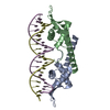
| ||||||||||
|---|---|---|---|---|---|---|---|---|---|---|---|
| 1 |
| ||||||||||
| Unit cell |
| ||||||||||
| Details | The whole biological assembly is contained in the asymmetric unit. It comprises a homodimer constructed from chain A and B bound to double stranded DNA (chains D and E). |
-Components
| #1: DNA chain | Mass: 6445.209 Da / Num. of mol.: 1 / Source method: obtained synthetically Details: PSEUDOSYMMETRIC B-SITE OF THE TERI SEQUENCE OF B. SUBTILIS | ||
|---|---|---|---|
| #2: DNA chain | Mass: 6436.195 Da / Num. of mol.: 1 / Source method: obtained synthetically Details: PSEUDOSYMMETRIC B-SITE OF THE TERI SEQUENCE OF B. SUBTILIS | ||
| #3: Protein | Mass: 14531.099 Da / Num. of mol.: 2 / Mutation: C110S Source method: isolated from a genetically manipulated source Source: (gene. exp.) #4: Water | ChemComp-HOH / |  Mass: 18.015 Da / Num. of mol.: 96 / Source method: isolated from a natural source / Formula: H2O Mass: 18.015 Da / Num. of mol.: 96 / Source method: isolated from a natural source / Formula: H2O |
-Experimental details
-Experiment
| Experiment | Method: X-RAY DIFFRACTION / Number of used crystals: 1 |
|---|
- Sample preparation
| Crystal | Density Matthews: 2.94 Å3/Da / Density % sol: 57.91 % | ||||||||||||||||||||
|---|---|---|---|---|---|---|---|---|---|---|---|---|---|---|---|---|---|---|---|---|---|
| Crystal grow | Temperature: 298 K / Method: vapor diffusion, hanging drop / pH: 4.6 Details: 5% PEG 4000, 0.1M sodium acetate, pH 4.6, VAPOR DIFFUSION, HANGING DROP at 298K | ||||||||||||||||||||
| Components of the solutions |
| ||||||||||||||||||||
| Crystal grow | *PLUS Temperature: 21 ℃ | ||||||||||||||||||||
| Components of the solutions | *PLUS
|
-Data collection
| Diffraction | Mean temperature: 100 K |
|---|---|
| Diffraction source | Source: SYNCHROTRON / Site: APS  / Beamline: 14-BM-C / Wavelength: 1 / Beamline: 14-BM-C / Wavelength: 1 |
| Detector | Type: ADSC QUANTUM 4 / Detector: CCD / Date: Dec 16, 1999 |
| Radiation | Protocol: SINGLE WAVELENGTH / Monochromatic (M) / Laue (L): M / Scattering type: x-ray |
| Radiation wavelength | Wavelength: 1 Å / Relative weight: 1 |
| Reflection | Resolution: 2.5→50 Å / Num. all: 15166 / % possible obs: 87.9 % / Observed criterion σ(I): -3 / Redundancy: 9.7 % / Biso Wilson estimate: 60.5 Å2 / Rmerge(I) obs: 0.073 / Net I/σ(I): 13.7 |
| Reflection shell | Resolution: 2.5→2.58 Å / Redundancy: 5.8 % / Rmerge(I) obs: 0.175 / % possible all: 54.2 |
| Reflection | *PLUS Lowest resolution: 50 Å / Num. obs: 15166 / Num. measured all: 147110 |
| Reflection shell | *PLUS % possible obs: 54.2 % |
- Processing
| Software |
| |||||||||||||||||||||||||
|---|---|---|---|---|---|---|---|---|---|---|---|---|---|---|---|---|---|---|---|---|---|---|---|---|---|---|
| Refinement | Resolution: 2.5→50 Å / Cross valid method: THROUGHOUT / σ(F): 0 / σ(I): 0 Stereochemistry target values: protein_rep.param, dna-rna_rep.param, water_rep.param
| |||||||||||||||||||||||||
| Displacement parameters |
| |||||||||||||||||||||||||
| Refinement step | Cycle: LAST / Resolution: 2.5→50 Å
| |||||||||||||||||||||||||
| Refine LS restraints |
| |||||||||||||||||||||||||
| Software | *PLUS Name: CNS / Classification: refinement | |||||||||||||||||||||||||
| Refinement | *PLUS Highest resolution: 2.5 Å / Lowest resolution: 50 Å / σ(F): 0 / % reflection Rfree: 10 % / Rfactor obs: 0.232 | |||||||||||||||||||||||||
| Solvent computation | *PLUS | |||||||||||||||||||||||||
| Displacement parameters | *PLUS | |||||||||||||||||||||||||
| LS refinement shell | *PLUS Highest resolution: 2.5 Å / Lowest resolution: 2.59 Å / Rfactor Rfree: 0.381 / Rfactor obs: 0.301 |
