 Movie
Movie Controller

[English] 日本語
 Yorodumi
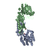
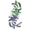
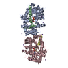
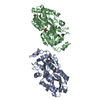
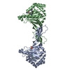
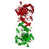
- PDB-5vo3: Crystal structure of DapE in complex with the products (succinic ... -
+ Open data

- Basic information
| Entry | Database: PDB / ID: 5vo3 | |||||||||
|---|---|---|---|---|---|---|---|---|---|---|
| Title | Crystal structure of DapE in complex with the products (succinic acid and diaminopimelic acid) | |||||||||
 Components Components | Succinyl-diaminopimelate desuccinylase | |||||||||
 Keywords Keywords | HYDROLASE / DapE / hydrolaze / Succinic acid / diaminopimelic acid / M28 / Structural Genomics / PSI-Biology | |||||||||
| Function / homology |  Function and homology information Function and homology informationsuccinyl-diaminopimelate desuccinylase / succinyl-diaminopimelate desuccinylase activity / : / : / cobalt ion binding / zinc ion binding / cytosol Similarity search - Function | |||||||||
| Biological species |  Haemophilus influenzae (bacteria) Haemophilus influenzae (bacteria) | |||||||||
| Method |  X-RAY DIFFRACTION / SYNCHROTRON / MOLECULAR REPLACEMENT / Resolution: 1.954 Å X-RAY DIFFRACTION / SYNCHROTRON / MOLECULAR REPLACEMENT / Resolution: 1.954 Å | |||||||||
 Authors Authors | Nocek, B. | |||||||||
 Citation Citation | Journal: Biochemistry / Year: 2018 Title: Structural Evidence of a Major Conformational Change Triggered by Substrate Binding in DapE Enzymes: Impact on the Catalytic Mechanism. Authors: Nocek, B. / Reidl, C. / Starus, A. / Heath, T. / Bienvenue, D. / Osipiuk, J. / Jedrzejczak, R. / Joachimiak, A. / Becker, D.P. / Holz, R.C. | |||||||||
| History |
|
- Structure visualization
| Structure viewer | Molecule: MolmilJmol/JSmol |
|---|
- Downloads & links
-Download
| PDBx/mmCIF format | 5vo3.cif.gz | 229.2 KB | Display | PDBx/mmCIF format |
|---|---|---|---|---|
| PDB format | pdb5vo3.ent.gz | 186.1 KB | Display | PDB format |
| PDBx/mmJSON format | 5vo3.json.gz | Tree view | PDBx/mmJSON format | |
| Others |  Other downloads Other downloads |
-Validation report
| Arichive directory | https://data.pdbj.org/pub/pdb/validation_reports/vo/5vo3ftp://data.pdbj.org/pub/pdb/validation_reports/vo/5vo3 | HTTPS FTP |
|---|
-Related structure data
| Related structure data | 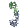 3iszS S: Starting model for refinement |
|---|---|
| Similar structure data |


-Links
 PDBj
PDBj
- Assembly
| Deposited unit | 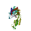
| |||||||||
|---|---|---|---|---|---|---|---|---|---|---|
| 1 | 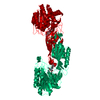
| |||||||||
| Unit cell |
| |||||||||
| Components on special symmetry positions |
|
-Components
| #1: Protein | Mass: 41654.211 Da / Num. of mol.: 1 Source method: isolated from a genetically manipulated source Source: (gene. exp.) Haemophilus influenzae (bacteria) / Gene: dapE, HI_0102Production host: References: UniProt: P44514, succinyl-diaminopimelate desuccinylase | ||
|---|---|---|---|
| #2: Chemical | ChemComp-SIN /   Mass: 118.088 Da / Num. of mol.: 1 Mass: 118.088 Da / Num. of mol.: 1Source method: isolated from a genetically manipulated source Formula: C4H6O4 | ||
| #3: Chemical | ChemComp-API / 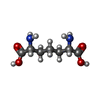  Type: L-peptide linking / Mass: 190.197 Da / Num. of mol.: 1 Type: L-peptide linking / Mass: 190.197 Da / Num. of mol.: 1Source method: isolated from a genetically manipulated source Formula: C7H14N2O4 | ||
| #4: Chemical |   Mass: 65.409 Da / Num. of mol.: 2 Mass: 65.409 Da / Num. of mol.: 2Source method: isolated from a genetically manipulated source Formula: Zn #5: Water | ChemComp-HOH / |  Mass: 18.015 Da / Num. of mol.: 229 / Source method: isolated from a natural source / Formula: H2O Mass: 18.015 Da / Num. of mol.: 229 / Source method: isolated from a natural source / Formula: H2O |
-Experimental details
-Experiment
| Experiment | Method: X-RAY DIFFRACTION / Number of used crystals: 1 |
|---|
- Sample preparation
| Crystal | Density Matthews: 3.43 Å3/Da / Density % sol: 64.12 % |
|---|---|
| Crystal grow | Temperature: 293 K / Method: vapor diffusion, sitting drop / pH: 7.5 Details: 0.15 M Hepes pH 7.5, 0.06 M Sodium potassium tartrate, 26 % Peg 8000 |
-Data collection
| Diffraction | Mean temperature: 100 K |
|---|---|
| Diffraction source | Source: SYNCHROTRON / Site: APS  / Beamline: 19-ID / Wavelength: 0.9794 Å / Beamline: 19-ID / Wavelength: 0.9794 Å |
| Detector | Type: ADSC QUANTUM 315r / Detector: CCD / Date: Feb 8, 2016 / Details: mirrors |
| Radiation | Monochromator: double crystals / Protocol: SINGLE WAVELENGTH / Monochromatic (M) / Laue (L): M / Scattering type: x-ray |
| Radiation wavelength | Wavelength: 0.9794 Å / Relative weight: 1 |
| Reflection | Resolution: 1.95→40 Å / Num. obs: 60902 / % possible obs: 99.5 % / Redundancy: 4 % / Rmerge(I) obs: 0.073 / Net I/σ(I): 16 |
| Reflection shell | Resolution: 1.9539→1.9844 Å |
- Processing
| Software |
| |||||||||||||||||||||||||||||||||||||||||||||||||||||||||||||||||||||||||||||||||||||||||||||||||||||||||||||||||||||||||||||||||||||||||||||||||||||||||||||||||
|---|---|---|---|---|---|---|---|---|---|---|---|---|---|---|---|---|---|---|---|---|---|---|---|---|---|---|---|---|---|---|---|---|---|---|---|---|---|---|---|---|---|---|---|---|---|---|---|---|---|---|---|---|---|---|---|---|---|---|---|---|---|---|---|---|---|---|---|---|---|---|---|---|---|---|---|---|---|---|---|---|---|---|---|---|---|---|---|---|---|---|---|---|---|---|---|---|---|---|---|---|---|---|---|---|---|---|---|---|---|---|---|---|---|---|---|---|---|---|---|---|---|---|---|---|---|---|---|---|---|---|---|---|---|---|---|---|---|---|---|---|---|---|---|---|---|---|---|---|---|---|---|---|---|---|---|---|---|---|---|---|---|---|
| Refinement | Method to determine structure: MOLECULAR REPLACEMENT Starting model: 3ISZ Resolution: 1.954→35.321 Å / SU ML: 0.14 / Cross valid method: FREE R-VALUE / σ(F): 1.35 / Phase error: 19.35
| |||||||||||||||||||||||||||||||||||||||||||||||||||||||||||||||||||||||||||||||||||||||||||||||||||||||||||||||||||||||||||||||||||||||||||||||||||||||||||||||||
| Solvent computation | Shrinkage radii: 0.9 Å / VDW probe radii: 1.11 Å | |||||||||||||||||||||||||||||||||||||||||||||||||||||||||||||||||||||||||||||||||||||||||||||||||||||||||||||||||||||||||||||||||||||||||||||||||||||||||||||||||
| Refinement step | Cycle: LAST / Resolution: 1.954→35.321 Å
| |||||||||||||||||||||||||||||||||||||||||||||||||||||||||||||||||||||||||||||||||||||||||||||||||||||||||||||||||||||||||||||||||||||||||||||||||||||||||||||||||
| Refine LS restraints |
| |||||||||||||||||||||||||||||||||||||||||||||||||||||||||||||||||||||||||||||||||||||||||||||||||||||||||||||||||||||||||||||||||||||||||||||||||||||||||||||||||
| LS refinement shell |
| |||||||||||||||||||||||||||||||||||||||||||||||||||||||||||||||||||||||||||||||||||||||||||||||||||||||||||||||||||||||||||||||||||||||||||||||||||||||||||||||||
| Refinement TLS params. | Method: refined / Refine-ID: X-RAY DIFFRACTION
| |||||||||||||||||||||||||||||||||||||||||||||||||||||||||||||||||||||||||||||||||||||||||||||||||||||||||||||||||||||||||||||||||||||||||||||||||||||||||||||||||
| Refinement TLS group |
|
