 Movie
Movie Controller

[English] 日本語
 Yorodumi
- PDB-1kjy: Crystal Structure of Human G[alpha]i1 Bound to the GoLoco Motif o... -
+ Open data

- Basic information
| Entry | Database: PDB / ID: 1kjy | ||||||
|---|---|---|---|---|---|---|---|
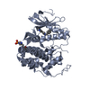
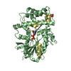
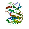
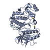
| Title | Crystal Structure of Human G[alpha]i1 Bound to the GoLoco Motif of RGS14 | ||||||
 Components Components |
| ||||||
 Keywords Keywords | SIGNALING PROTEIN / Protein-peptide complex | ||||||
| Function / homology |  Function and homology information Function and homology informationzygote asymmetric cell division / negative regulation of synaptic plasticity / regulation of G protein-coupled receptor signaling pathway / GTPase activating protein binding / G alpha (i) signalling events / spindle organization / GDP-dissociation inhibitor activity / nucleocytoplasmic transport / negative regulation of G protein-coupled receptor signaling pathway / positive regulation of neurogenesis ...zygote asymmetric cell division / negative regulation of synaptic plasticity / regulation of G protein-coupled receptor signaling pathway / GTPase activating protein binding / G alpha (i) signalling events / spindle organization / GDP-dissociation inhibitor activity / nucleocytoplasmic transport / negative regulation of G protein-coupled receptor signaling pathway / positive regulation of neurogenesis / negative regulation of MAP kinase activity / platelet-derived growth factor receptor signaling pathway / G-protein alpha-subunit binding / long-term memory / adenylate cyclase inhibitor activity / positive regulation of protein localization to cell cortex / T cell migration / positive regulation of relaxation of smooth muscle / Adenylate cyclase inhibitory pathway / D2 dopamine receptor binding / adenylate cyclase-inhibiting serotonin receptor signaling pathway / G protein-coupled serotonin receptor binding / cellular response to forskolin / GTPase activator activity / regulation of mitotic spindle organization / chemokine-mediated signaling pathway / learning / chromosome segregation / Regulation of insulin secretion / neuropeptide signaling pathway / response to prostaglandin E / negative regulation of ERK1 and ERK2 cascade / positive regulation of cholesterol biosynthetic process / PML body / negative regulation of insulin secretion / visual learning / G protein-coupled receptor binding / modulation of chemical synaptic transmission / response to peptide hormone / spindle / centriolar satellite / G-protein beta/gamma-subunit complex binding / spindle pole / adenylate cyclase-modulating G protein-coupled receptor signaling pathway / adenylate cyclase-inhibiting G protein-coupled receptor signaling pathway / ADP signalling through P2Y purinoceptor 12 / long-term synaptic potentiation / GDP binding / Adrenaline,noradrenaline inhibits insulin secretion / G alpha (z) signalling events / ADORA2B mediated anti-inflammatory cytokines production / mitotic cell cycle / GPER1 signaling / heterotrimeric G-protein complex / sperm principal piece / adenylate cyclase-activating G protein-coupled receptor signaling pathway / signaling receptor complex adaptor activity / response to oxidative stress / G protein activity / midbody / cell cortex / microtubule binding / G alpha (i) signalling events / dendritic spine / G alpha (s) signalling events / microtubule / Hydrolases; Acting on acid anhydrides; Acting on GTP to facilitate cellular and subcellular movement / Extra-nuclear estrogen signaling / postsynaptic density / nuclear body / ciliary basal body / G protein-coupled receptor signaling pathway / cell division / lysosomal membrane / GTPase activity / centrosome / dendrite / protein kinase binding / GTP binding / nucleolus / glutamatergic synapse / magnesium ion binding / Golgi apparatus / extracellular exosome / nucleoplasm / nucleus / plasma membrane / cytoplasm / cytosol Similarity search - Function | ||||||
| Biological species |  Homo sapiens (human) Homo sapiens (human) | ||||||
| Method |  X-RAY DIFFRACTION / MOLECULAR REPLACEMENT / Resolution: 2.7 Å X-RAY DIFFRACTION / MOLECULAR REPLACEMENT / Resolution: 2.7 Å | ||||||
 Authors Authors | Kimple, R.J. / Kimple, M.E. / Betts, L. / Sondek, J. / Siderovski, D.P. | ||||||
 Citation Citation | Journal: Nature / Year: 2002 Title: Structural determinants for GoLoco-induced inhibition of nucleotide release by Galpha subunits. Authors: Kimple, R.J. / Kimple, M.E. / Betts, L. / Sondek, J. / Siderovski, D.P. | ||||||
| History |
| ||||||
| Remark 999 | SEQUENCE THE AUTHORS MAINTAIN THAT THEIR SEQUENCE IS CORRECT AND THAT RESIDUE 30 IS GLY FOR CHAINS A & B. |
- Structure visualization
| Structure viewer | Molecule: MolmilJmol/JSmol |
|---|
- Downloads & links
-Download
| PDBx/mmCIF format | 1kjy.cif.gz | 162.8 KB | Display | PDBx/mmCIF format |
|---|---|---|---|---|
| PDB format | pdb1kjy.ent.gz | 125 KB | Display | PDB format |
| PDBx/mmJSON format | 1kjy.json.gz | Tree view | PDBx/mmJSON format | |
| Others |  Other downloads Other downloads |
-Validation report
| Arichive directory | https://data.pdbj.org/pub/pdb/validation_reports/kj/1kjyftp://data.pdbj.org/pub/pdb/validation_reports/kj/1kjy | HTTPS FTP |
|---|
-Related structure data
| Related structure data |  1bofS S: Starting model for refinement |
|---|---|
| Similar structure data |


-Links
 PDBj
PDBj


- Assembly
| Deposited unit | 
| ||||||||
|---|---|---|---|---|---|---|---|---|---|
| 1 | 
| ||||||||
| 2 | 
| ||||||||
| 3 | 
| ||||||||
| Unit cell |
| ||||||||
| Components on special symmetry positions |
|
-Components
-Protein / Protein/peptide , 2 types, 4 molecules ACBD
| #1: Protein | Mass: 37159.348 Da / Num. of mol.: 2 Source method: isolated from a genetically manipulated source Source: (gene. exp.) Homo sapiens (human) / Plasmid: pPROexHTb / Species (production host): Escherichia coli / Production host:  #2: Protein/peptide | Mass: 4093.621 Da / Num. of mol.: 2 / Source method: obtained synthetically Details: The peptide was chemically synthesized. The sequence of the peptide is naturally found in Rattus norvegicus (rat). References: UniProt: O08773 |
|---|
-Non-polymers , 4 types, 225 molecules 


| #3: Chemical | ChemComp-CS /  Mass: 132.905 Da / Num. of mol.: 15 / Source method: obtained synthetically / Formula: Cs Mass: 132.905 Da / Num. of mol.: 15 / Source method: obtained synthetically / Formula: Cs#4: Chemical |  Type: RNA linking / Mass: 443.201 Da / Num. of mol.: 2 / Source method: obtained synthetically / Formula: C10H15N5O11P2 / Comment: GDP, energy-carrying molecule*YM Type: RNA linking / Mass: 443.201 Da / Num. of mol.: 2 / Source method: obtained synthetically / Formula: C10H15N5O11P2 / Comment: GDP, energy-carrying molecule*YM#5: Chemical |  Mass: 24.305 Da / Num. of mol.: 2 / Source method: obtained synthetically / Formula: Mg Mass: 24.305 Da / Num. of mol.: 2 / Source method: obtained synthetically / Formula: Mg#6: Water | ChemComp-HOH / | Mass: 18.015 Da / Num. of mol.: 206 / Source method: isolated from a natural source / Formula: H2O |
|---|
-Experimental details
-Experiment
| Experiment | Method: X-RAY DIFFRACTION / Number of used crystals: 1 |
|---|
- Sample preparation
| Crystal | Density Matthews: 3.23 Å3/Da / Density % sol: 61.98 % | |||||||||||||||||||||||||||||||||||||||||||||||||||||||||||||||
|---|---|---|---|---|---|---|---|---|---|---|---|---|---|---|---|---|---|---|---|---|---|---|---|---|---|---|---|---|---|---|---|---|---|---|---|---|---|---|---|---|---|---|---|---|---|---|---|---|---|---|---|---|---|---|---|---|---|---|---|---|---|---|---|---|
| Crystal grow | Temperature: 291 K / Method: vapor diffusion, sitting drop / pH: 4.6 Details: Cesium Sulfate, sodium acetate, glycerol, pH 4.6, VAPOR DIFFUSION, SITTING DROP, temperature 291K | |||||||||||||||||||||||||||||||||||||||||||||||||||||||||||||||
| Crystal grow | *PLUS Temperature: 18 ℃ / pH: 7.5 | |||||||||||||||||||||||||||||||||||||||||||||||||||||||||||||||
| Components of the solutions | *PLUS
|
-Data collection
| Diffraction | Mean temperature: 100 K |
|---|---|
| Diffraction source | Source: ROTATING ANODE / Type: RIGAKU RUH3R / Wavelength: 1.5418 Å |
| Detector | Type: RIGAKU RAXIS IV++ / Detector: IMAGE PLATE / Date: Jul 17, 2001 / Details: Osmic Confocal Blue |
| Radiation | Monochromator: Ni Filter / Protocol: SINGLE WAVELENGTH / Monochromatic (M) / Laue (L): M / Scattering type: x-ray |
| Radiation wavelength | Wavelength: 1.5418 Å / Relative weight: 1 |
| Reflection | Resolution: 2.7→20 Å / Num. obs: 136468 / % possible obs: 99.1 % / Observed criterion σ(I): 2.5 / Redundancy: 4.6 % / Biso Wilson estimate: 39.9 Å2 / Rmerge(I) obs: 0.111 / Rsym value: 0.088 / Net I/σ(I): 14 |
| Reflection shell | Resolution: 2.7→2.8 Å / Rmerge(I) obs: 0.48 / Mean I/σ(I) obs: 3.4 / Num. unique all: 2875 / Rsym value: 0.45 / % possible all: 97.5 |
| Reflection | *PLUS Lowest resolution: 20 Å / Num. obs: 29900 / Num. measured all: 136468 / Rmerge(I) obs: 0.111 |
| Reflection shell | *PLUS % possible obs: 97.5 % / Num. unique obs: 2875 / Rmerge(I) obs: 0.48 |
- Processing
| Software |
| ||||||||||||||||||||||||||||||||||||||||||||||||||||||||||||
|---|---|---|---|---|---|---|---|---|---|---|---|---|---|---|---|---|---|---|---|---|---|---|---|---|---|---|---|---|---|---|---|---|---|---|---|---|---|---|---|---|---|---|---|---|---|---|---|---|---|---|---|---|---|---|---|---|---|---|---|---|---|
| Refinement | Method to determine structure: MOLECULAR REPLACEMENT Starting model: PDB entry 1BOF Resolution: 2.7→20 Å / Rfactor Rfree error: 0.008 / Data cutoff high absF: 1706201.14 / Data cutoff low absF: 0 / Isotropic thermal model: RESTRAINED / Cross valid method: THROUGHOUT / σ(F): 0 / Stereochemistry target values: Engh and Huber
| ||||||||||||||||||||||||||||||||||||||||||||||||||||||||||||
| Solvent computation | Solvent model: FLAT MODEL / Bsol: 39.227 Å2 / ksol: 0.340631 e/Å3 | ||||||||||||||||||||||||||||||||||||||||||||||||||||||||||||
| Displacement parameters | Biso mean: 42 Å2
| ||||||||||||||||||||||||||||||||||||||||||||||||||||||||||||
| Refine analyze |
| ||||||||||||||||||||||||||||||||||||||||||||||||||||||||||||
| Refinement step | Cycle: LAST / Resolution: 2.7→20 Å
| ||||||||||||||||||||||||||||||||||||||||||||||||||||||||||||
| Refine LS restraints |
| ||||||||||||||||||||||||||||||||||||||||||||||||||||||||||||
| LS refinement shell | Resolution: 2.7→2.87 Å / Rfactor Rfree error: 0.025 / Total num. of bins used: 6
| ||||||||||||||||||||||||||||||||||||||||||||||||||||||||||||
| Xplor file |
| ||||||||||||||||||||||||||||||||||||||||||||||||||||||||||||
| Refinement | *PLUS Lowest resolution: 20 Å / Rfactor obs: 0.238 / Rfactor Rfree: 0.299 / Rfactor Rwork: 0.238 | ||||||||||||||||||||||||||||||||||||||||||||||||||||||||||||
| Solvent computation | *PLUS | ||||||||||||||||||||||||||||||||||||||||||||||||||||||||||||
| Displacement parameters | *PLUS | ||||||||||||||||||||||||||||||||||||||||||||||||||||||||||||
| Refine LS restraints | *PLUS
| ||||||||||||||||||||||||||||||||||||||||||||||||||||||||||||
| LS refinement shell | *PLUS Highest resolution: 2.7 Å / Rfactor Rfree: 0.383 / Rfactor Rwork: 0.313 / Rfactor obs: 0.313 |
