 Movie
Movie Controller

[English] 日本語
 Yorodumi
- PDB-3qi2: A Galpha P-loop mutation prevents transition to the activated sta... -
+ Open data

- Basic information
| Entry | Database: PDB / ID: 3qi2 | ||||||
|---|---|---|---|---|---|---|---|
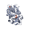
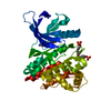
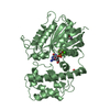
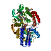
| Title | A Galpha P-loop mutation prevents transition to the activated state: G42R bound to RGS14 GoLoco | ||||||
 Components Components |
| ||||||
 Keywords Keywords | SIGNALING PROTEIN / RGS14 GoLoco / Ras-like domain / all-helical domain / GoLoco motif / arginine finger / lipoprotein / transducer / guanine nucleotide dissociation inhibitor / GTP binding / nucleotide binding / ADP-ribosylation | ||||||
| Function / homology |  Function and homology information Function and homology informationzygote asymmetric cell division / negative regulation of synaptic plasticity / regulation of G protein-coupled receptor signaling pathway / GTPase activating protein binding / spindle organization / GDP-dissociation inhibitor activity / nucleocytoplasmic transport / negative regulation of G protein-coupled receptor signaling pathway / positive regulation of neurogenesis / negative regulation of MAP kinase activity ...zygote asymmetric cell division / negative regulation of synaptic plasticity / regulation of G protein-coupled receptor signaling pathway / GTPase activating protein binding / spindle organization / GDP-dissociation inhibitor activity / nucleocytoplasmic transport / negative regulation of G protein-coupled receptor signaling pathway / positive regulation of neurogenesis / negative regulation of MAP kinase activity / platelet-derived growth factor receptor signaling pathway / G-protein alpha-subunit binding / long-term memory / adenylate cyclase inhibitor activity / positive regulation of protein localization to cell cortex / T cell migration / positive regulation of relaxation of smooth muscle / Adenylate cyclase inhibitory pathway / D2 dopamine receptor binding / adenylate cyclase-inhibiting serotonin receptor signaling pathway / G protein-coupled serotonin receptor binding / cellular response to forskolin / regulation of mitotic spindle organization / GTPase activator activity / chemokine-mediated signaling pathway / learning / chromosome segregation / Regulation of insulin secretion / neuropeptide signaling pathway / response to prostaglandin E / negative regulation of ERK1 and ERK2 cascade / positive regulation of cholesterol biosynthetic process / PML body / negative regulation of insulin secretion / visual learning / G protein-coupled receptor binding / response to peptide hormone / spindle / centriolar satellite / G-protein beta/gamma-subunit complex binding / spindle pole / adenylate cyclase-modulating G protein-coupled receptor signaling pathway / adenylate cyclase-inhibiting G protein-coupled receptor signaling pathway / ADP signalling through P2Y purinoceptor 12 / GDP binding / long-term synaptic potentiation / Adrenaline,noradrenaline inhibits insulin secretion / G alpha (z) signalling events / ADORA2B mediated anti-inflammatory cytokines production / mitotic cell cycle / GPER1 signaling / heterotrimeric G-protein complex / sperm principal piece / adenylate cyclase-activating G protein-coupled receptor signaling pathway / signaling receptor complex adaptor activity / response to oxidative stress / G protein activity / midbody / cell cortex / microtubule binding / G alpha (i) signalling events / G alpha (s) signalling events / dendritic spine / microtubule / Hydrolases; Acting on acid anhydrides; Acting on GTP to facilitate cellular and subcellular movement / Extra-nuclear estrogen signaling / postsynaptic density / nuclear body / ciliary basal body / G protein-coupled receptor signaling pathway / cell division / lysosomal membrane / GTPase activity / centrosome / dendrite / protein kinase binding / GTP binding / nucleolus / glutamatergic synapse / magnesium ion binding / Golgi apparatus / extracellular exosome / nucleoplasm / nucleus / plasma membrane / cytoplasm / cytosol Similarity search - Function | ||||||
| Biological species |  Homo sapiens (human) Homo sapiens (human) | ||||||
| Method |  X-RAY DIFFRACTION / SYNCHROTRON / MOLECULAR REPLACEMENT / Resolution: 2.797 Å X-RAY DIFFRACTION / SYNCHROTRON / MOLECULAR REPLACEMENT / Resolution: 2.797 Å | ||||||
 Authors Authors | Bosch, D.E. / Willard, F.S. / Kimple, A.J. / Miley, M.J. / Siderovski, D.P. | ||||||
 Citation Citation | Journal: Plos Pathog. / Year: 2012 Title: A P-loop Mutation in Galpha Subunits Prevents Transition to the Active State: Implications for G-protein Signaling in Fungal Pathogenesis Authors: Bosch, D.E. / Willard, F.S. / Ramanujam, R. / Kimple, A.J. / Willard, M.D. / Naqvi, N.I. / Siderovski, D.P. #1: Journal: J.Mol.Biol. / Year: 2007Title: Structure-based protocol for identifying mutations that enhance protein-protein binding affinities. Authors: Sammond, D.W. / Eletr, Z.M. / Purbeck, C. / Kimple, R.J. / Siderovski, D.P. / Kuhlman, B. #2: Journal: Nature / Year: 2002Title: Structural determinants for GoLoco-induced inhibition of nucleotide release by Galpha subunits. Authors: Kimple, R.J. / Kimple, M.E. / Betts, L. / Sondek, J. / Siderovski, D.P. | ||||||
| History |
|
- Structure visualization
| Structure viewer | Molecule: MolmilJmol/JSmol |
|---|
- Downloads & links
-Download
| PDBx/mmCIF format | 3qi2.cif.gz | 283.8 KB | Display | PDBx/mmCIF format |
|---|---|---|---|---|
| PDB format | pdb3qi2.ent.gz | 230.8 KB | Display | PDB format |
| PDBx/mmJSON format | 3qi2.json.gz | Tree view | PDBx/mmJSON format | |
| Others |  Other downloads Other downloads |
-Validation report
| Arichive directory | https://data.pdbj.org/pub/pdb/validation_reports/qi/3qi2ftp://data.pdbj.org/pub/pdb/validation_reports/qi/3qi2 | HTTPS FTP |
|---|
-Related structure data
| Related structure data |  3qe0C  2om2S C: citing same article ( S: Starting model for refinement |
|---|---|
| Similar structure data |

-Links
 PDBj
PDBj


- Assembly
| Deposited unit | 
| ||||||||
|---|---|---|---|---|---|---|---|---|---|
| 1 | 
| ||||||||
| 2 | 
| ||||||||
| Unit cell |
|
-Components
-Protein / Protein/peptide , 2 types, 4 molecules ABCD
| #1: Protein | Mass: 37531.750 Da / Num. of mol.: 2 / Fragment: alpha-i1 subunit, residues 31-354 / Mutation: G42R Source method: isolated from a genetically manipulated source Source: (gene. exp.) Homo sapiens (human) / Gene: GNAI1 / Plasmid: pLIC-His / Production host:  #2: Protein/peptide | Mass: 4093.621 Da / Num. of mol.: 2 / Fragment: GoLoco motif peptide, residues 497-532 / Source method: obtained synthetically Details: synthetic GoLoco motif peptide identical to human RGS14 497-532 References: UniProt: O43566 |
|---|
-Non-polymers , 4 types, 42 molecules 


| #3: Chemical |  Type: RNA linking / Mass: 443.201 Da / Num. of mol.: 2 / Source method: obtained synthetically / Formula: C10H15N5O11P2 / Comment: GDP, energy-carrying molecule*YM Type: RNA linking / Mass: 443.201 Da / Num. of mol.: 2 / Source method: obtained synthetically / Formula: C10H15N5O11P2 / Comment: GDP, energy-carrying molecule*YM#4: Chemical |  Mass: 96.063 Da / Num. of mol.: 2 / Source method: obtained synthetically / Formula: SO4 Mass: 96.063 Da / Num. of mol.: 2 / Source method: obtained synthetically / Formula: SO4#5: Chemical | ChemComp-GOL / |  Mass: 92.094 Da / Num. of mol.: 1 / Source method: obtained synthetically / Formula: C3H8O3 Mass: 92.094 Da / Num. of mol.: 1 / Source method: obtained synthetically / Formula: C3H8O3#6: Water | ChemComp-HOH / | Mass: 18.015 Da / Num. of mol.: 37 / Source method: isolated from a natural source / Formula: H2O |
|---|
-Experimental details
-Experiment
| Experiment | Method: X-RAY DIFFRACTION / Number of used crystals: 1 |
|---|
- Sample preparation
| Crystal | Density Matthews: 2.8 Å3/Da / Density % sol: 56.07 % |
|---|---|
| Crystal grow | Temperature: 291 K / Method: vapor diffusion, hanging drop / pH: 5 Details: Hanging drops were a 1:1 mixture of protein-peptide complex in buffer (10 mM Tris pH 7.5, 1 mM magnesium chloride, 5% (w/v) glycerol, 5 mM DTT) and well solution (1.7 M ammonium sulfate, 100 ...Details: Hanging drops were a 1:1 mixture of protein-peptide complex in buffer (10 mM Tris pH 7.5, 1 mM magnesium chloride, 5% (w/v) glycerol, 5 mM DTT) and well solution (1.7 M ammonium sulfate, 100 mM sodium acetate pH 5.0, 200 mM magnesium chloride, 10% (w/v) glycerol), VAPOR DIFFUSION, HANGING DROP, temperature 291K |
-Data collection
| Diffraction | Mean temperature: 100 K |
|---|---|
| Diffraction source | Source: SYNCHROTRON / Site: APS  / Beamline: 22-ID / Wavelength: 1 Å / Beamline: 22-ID / Wavelength: 1 Å |
| Detector | Type: MAR scanner 300 mm plate / Detector: IMAGE PLATE / Date: Nov 1, 2010 / Details: custom |
| Radiation | Protocol: SINGLE WAVELENGTH / Monochromatic (M) / Laue (L): M / Scattering type: x-ray |
| Radiation wavelength | Wavelength: 1 Å / Relative weight: 1 |
| Reflection | Resolution: 2.797→29.705 Å / Num. all: 23542 / Num. obs: 18462 / % possible obs: 78.42 % / Observed criterion σ(I): -3 / Biso Wilson estimate: 51.89 Å2 / Net I/σ(I): 2 |
| Reflection shell | Resolution: 2.797→2.944 Å / Mean I/σ(I) obs: 2 / % possible all: 31 |
- Processing
| Software |
| |||||||||||||||||||||||||||||||||||||||||||||||||||||||||||||||||||||||||||||||||||||||||||||||||||||||||||||||||||||||||||||||||||||||||||||||||||||||||||||||||||||||||||||||||||||||||||||||||||||||||||||||||||||||||||||||||||||||||||||||||||||||||||||||||||||||||||||||||||
|---|---|---|---|---|---|---|---|---|---|---|---|---|---|---|---|---|---|---|---|---|---|---|---|---|---|---|---|---|---|---|---|---|---|---|---|---|---|---|---|---|---|---|---|---|---|---|---|---|---|---|---|---|---|---|---|---|---|---|---|---|---|---|---|---|---|---|---|---|---|---|---|---|---|---|---|---|---|---|---|---|---|---|---|---|---|---|---|---|---|---|---|---|---|---|---|---|---|---|---|---|---|---|---|---|---|---|---|---|---|---|---|---|---|---|---|---|---|---|---|---|---|---|---|---|---|---|---|---|---|---|---|---|---|---|---|---|---|---|---|---|---|---|---|---|---|---|---|---|---|---|---|---|---|---|---|---|---|---|---|---|---|---|---|---|---|---|---|---|---|---|---|---|---|---|---|---|---|---|---|---|---|---|---|---|---|---|---|---|---|---|---|---|---|---|---|---|---|---|---|---|---|---|---|---|---|---|---|---|---|---|---|---|---|---|---|---|---|---|---|---|---|---|---|---|---|---|---|---|---|---|---|---|---|---|---|---|---|---|---|---|---|---|---|---|---|---|---|---|---|---|---|---|---|---|---|---|---|---|---|---|---|---|---|---|---|---|---|---|---|---|---|---|---|---|---|---|
| Refinement | Method to determine structure: MOLECULAR REPLACEMENT Starting model: 2om2 Resolution: 2.797→29.705 Å / SU ML: 0.41 / σ(F): 0.71 / Stereochemistry target values: ML
| |||||||||||||||||||||||||||||||||||||||||||||||||||||||||||||||||||||||||||||||||||||||||||||||||||||||||||||||||||||||||||||||||||||||||||||||||||||||||||||||||||||||||||||||||||||||||||||||||||||||||||||||||||||||||||||||||||||||||||||||||||||||||||||||||||||||||||||||||||
| Solvent computation | Shrinkage radii: 0.9 Å / VDW probe radii: 1.11 Å / Solvent model: FLAT BULK SOLVENT MODEL / Bsol: 36.758 Å2 / ksol: 0.318 e/Å3 | |||||||||||||||||||||||||||||||||||||||||||||||||||||||||||||||||||||||||||||||||||||||||||||||||||||||||||||||||||||||||||||||||||||||||||||||||||||||||||||||||||||||||||||||||||||||||||||||||||||||||||||||||||||||||||||||||||||||||||||||||||||||||||||||||||||||||||||||||||
| Displacement parameters |
| |||||||||||||||||||||||||||||||||||||||||||||||||||||||||||||||||||||||||||||||||||||||||||||||||||||||||||||||||||||||||||||||||||||||||||||||||||||||||||||||||||||||||||||||||||||||||||||||||||||||||||||||||||||||||||||||||||||||||||||||||||||||||||||||||||||||||||||||||||
| Refinement step | Cycle: LAST / Resolution: 2.797→29.705 Å
| |||||||||||||||||||||||||||||||||||||||||||||||||||||||||||||||||||||||||||||||||||||||||||||||||||||||||||||||||||||||||||||||||||||||||||||||||||||||||||||||||||||||||||||||||||||||||||||||||||||||||||||||||||||||||||||||||||||||||||||||||||||||||||||||||||||||||||||||||||
| Refine LS restraints |
| |||||||||||||||||||||||||||||||||||||||||||||||||||||||||||||||||||||||||||||||||||||||||||||||||||||||||||||||||||||||||||||||||||||||||||||||||||||||||||||||||||||||||||||||||||||||||||||||||||||||||||||||||||||||||||||||||||||||||||||||||||||||||||||||||||||||||||||||||||
| LS refinement shell |
| |||||||||||||||||||||||||||||||||||||||||||||||||||||||||||||||||||||||||||||||||||||||||||||||||||||||||||||||||||||||||||||||||||||||||||||||||||||||||||||||||||||||||||||||||||||||||||||||||||||||||||||||||||||||||||||||||||||||||||||||||||||||||||||||||||||||||||||||||||
| Refinement TLS params. | Method: refined / Refine-ID: X-RAY DIFFRACTION
| |||||||||||||||||||||||||||||||||||||||||||||||||||||||||||||||||||||||||||||||||||||||||||||||||||||||||||||||||||||||||||||||||||||||||||||||||||||||||||||||||||||||||||||||||||||||||||||||||||||||||||||||||||||||||||||||||||||||||||||||||||||||||||||||||||||||||||||||||||
| Refinement TLS group |
|
