 Movie
Movie Controller

[English] 日本語
 Yorodumi
- PDB-2xns: Crystal Structure Of Human G alpha i1 Bound To A Designed Helical... -
+ Open data

- Basic information
| Entry | Database: PDB / ID: 2xns | ||||||
|---|---|---|---|---|---|---|---|
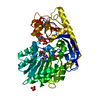
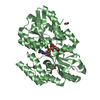
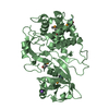
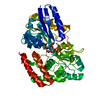
| Title | Crystal Structure Of Human G alpha i1 Bound To A Designed Helical Peptide Derived From The Goloco Motif Of RGS14 | ||||||
 Components Components |
| ||||||
 Keywords Keywords | HYDROLASE/PEPTIDE / HYDROLASE-PEPTIDE COMPLEX / ADP-RIBOSYLATION / ARGININE FINGER / GTP-BINDING / LIPOPROTEIN / NUCLEOTIDE-BINDING / PALMITATE / SIGNALING PROTEIN / TRANSDUCER / PROTEIN-PROTEIN INTERFACE DESIGN | ||||||
| Function / homology |  Function and homology information Function and homology informationzygote asymmetric cell division / negative regulation of synaptic plasticity / regulation of G protein-coupled receptor signaling pathway / GTPase activating protein binding / spindle organization / nucleocytoplasmic transport / GDP-dissociation inhibitor activity / negative regulation of G protein-coupled receptor signaling pathway / positive regulation of neurogenesis / platelet-derived growth factor receptor signaling pathway ...zygote asymmetric cell division / negative regulation of synaptic plasticity / regulation of G protein-coupled receptor signaling pathway / GTPase activating protein binding / spindle organization / nucleocytoplasmic transport / GDP-dissociation inhibitor activity / negative regulation of G protein-coupled receptor signaling pathway / positive regulation of neurogenesis / platelet-derived growth factor receptor signaling pathway / negative regulation of MAP kinase activity / G-protein alpha-subunit binding / long-term memory / regulation of eating behavior / adenylate cyclase inhibitor activity / positive regulation of protein localization to cell cortex / T cell migration / positive regulation of relaxation of smooth muscle / Adenylate cyclase inhibitory pathway / D2 dopamine receptor binding / adenylate cyclase-inhibiting serotonin receptor signaling pathway / G protein-coupled serotonin receptor binding / cellular response to forskolin / mast cell degranulation / GTPase activator activity / regulation of mitotic spindle organization / learning / chemokine-mediated signaling pathway / chromosome segregation / Regulation of insulin secretion / neuropeptide signaling pathway / response to prostaglandin E / negative regulation of ERK1 and ERK2 cascade / visual learning / positive regulation of cholesterol biosynthetic process / PML body / G protein-coupled receptor binding / response to peptide hormone / spindle / long-term synaptic potentiation / G-protein beta/gamma-subunit complex binding / spindle pole / adenylate cyclase-modulating G protein-coupled receptor signaling pathway / adenylate cyclase-inhibiting G protein-coupled receptor signaling pathway / GDP binding / ADP signalling through P2Y purinoceptor 12 / Adrenaline,noradrenaline inhibits insulin secretion / G alpha (z) signalling events / ADORA2B mediated anti-inflammatory cytokines production / mitotic cell cycle / GPER1 signaling / heterotrimeric G-protein complex / signaling receptor complex adaptor activity / adenylate cyclase-activating G protein-coupled receptor signaling pathway / response to oxidative stress / G protein activity / midbody / cell cortex / microtubule binding / G alpha (i) signalling events / dendritic spine / G alpha (s) signalling events / microtubule / Hydrolases; Acting on acid anhydrides; Acting on GTP to facilitate cellular and subcellular movement / Extra-nuclear estrogen signaling / ciliary basal body / postsynaptic density / nuclear body / G protein-coupled receptor signaling pathway / cell division / lysosomal membrane / GTPase activity / centrosome / dendrite / protein kinase binding / GTP binding / glutamatergic synapse / magnesium ion binding / extracellular exosome / nucleus / plasma membrane / cytosol / cytoplasm Similarity search - Function | ||||||
| Biological species |  HOMO SAPIENS (human) HOMO SAPIENS (human) | ||||||
| Method |  X-RAY DIFFRACTION / SYNCHROTRON / MOLECULAR REPLACEMENT / Resolution: 3.41 Å X-RAY DIFFRACTION / SYNCHROTRON / MOLECULAR REPLACEMENT / Resolution: 3.41 Å | ||||||
 Authors Authors | Bosch, D. / Sammond, D.W. / Butterfoss, G.L. / Machius, M. / Siderovski, D.P. / Kuhlman, B. | ||||||
 Citation Citation | Journal: J.Am.Chem.Soc. / Year: 2011 Title: Computational Design of the Sequence and Structure of a Protein-Binding Peptide. Authors: Sammond, D.W. / Bosch, D.E. / Butterfoss, G.L. / Purbeck, C. / Machius, M. / Siderovski, D.P. / Kuhlman, B. #1: Journal: J.Mol.Biol. / Year: 2007Title: Structure-Based Protocol for Identifying Mutations that Enhance Protein-Protein Binding Affinities. Authors: Sammond, D.W. / Eletr, Z.M. / Purbeck, C. / Kimple, R.J. / Siderovski, D.P. / Kuhlman, B. #2: Journal: Nature / Year: 2002Title: Structural Determinants for Goloco-Induced Inhibition of Nucleotide Release by Galpha Subunits. Authors: Kimple, R.J. / Kimple, M.E. / Betts, L. / Sondek, J. / Siderovski, D.P. | ||||||
| History |
|
- Structure visualization
| Structure viewer | Molecule: MolmilJmol/JSmol |
|---|
- Downloads & links
-Download
| PDBx/mmCIF format | 2xns.cif.gz | 308.9 KB | Display | PDBx/mmCIF format |
|---|---|---|---|---|
| PDB format | pdb2xns.ent.gz | 256.7 KB | Display | PDB format |
| PDBx/mmJSON format | 2xns.json.gz | Tree view | PDBx/mmJSON format | |
| Others |  Other downloads Other downloads |
-Validation report
| Arichive directory | https://data.pdbj.org/pub/pdb/validation_reports/xn/2xnsftp://data.pdbj.org/pub/pdb/validation_reports/xn/2xns | HTTPS FTP |
|---|
-Related structure data
| Related structure data |  2om2S S: Starting model for refinement |
|---|---|
| Similar structure data |


-Links
 PDBj
PDBj


- Assembly
| Deposited unit | 
| ||||||||||||||||||||||||||||||||||||||||||||||||||||||||||||
|---|---|---|---|---|---|---|---|---|---|---|---|---|---|---|---|---|---|---|---|---|---|---|---|---|---|---|---|---|---|---|---|---|---|---|---|---|---|---|---|---|---|---|---|---|---|---|---|---|---|---|---|---|---|---|---|---|---|---|---|---|---|
| 1 | 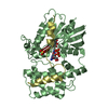
| ||||||||||||||||||||||||||||||||||||||||||||||||||||||||||||
| 2 | 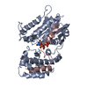
| ||||||||||||||||||||||||||||||||||||||||||||||||||||||||||||
| Unit cell |
| ||||||||||||||||||||||||||||||||||||||||||||||||||||||||||||
| Noncrystallographic symmetry (NCS) | NCS domain:
NCS domain segments:
NCS ensembles :
NCS oper:
|
-Components
-Protein / Protein/peptide , 2 types, 4 molecules ABCD
| #1: Protein | Mass: 37374.555 Da / Num. of mol.: 2 / Fragment: RESIDUES 30-354 Source method: isolated from a genetically manipulated source Source: (gene. exp.) HOMO SAPIENS (human) / Plasmid: PLICGNAI1 / Production host:  #2: Protein/peptide | Mass: 4695.173 Da / Num. of mol.: 2 / Fragment: RESIDUES 497-517 / Source method: obtained synthetically Details: DESIGNED HELICAL PEPTIDE DERIVED FROM REGULATOR OF G- PROTEIN SIGNALING 14 GOLOCO MOTIF PEPTIDE Source: (synth.) HOMO SAPIENS (human) / References: UniProt: O43566 |
|---|
-Non-polymers , 4 types, 16 molecules 


| #3: Chemical |  Type: RNA linking / Mass: 443.201 Da / Num. of mol.: 2 / Source method: obtained synthetically / Formula: C10H15N5O11P2 / Comment: GDP, energy-carrying molecule*YM Type: RNA linking / Mass: 443.201 Da / Num. of mol.: 2 / Source method: obtained synthetically / Formula: C10H15N5O11P2 / Comment: GDP, energy-carrying molecule*YM#4: Chemical |  Mass: 150.087 Da / Num. of mol.: 2 / Source method: obtained synthetically / Formula: C4H6O6 Mass: 150.087 Da / Num. of mol.: 2 / Source method: obtained synthetically / Formula: C4H6O6#5: Chemical |  Mass: 96.063 Da / Num. of mol.: 2 / Source method: obtained synthetically / Formula: SO4 Mass: 96.063 Da / Num. of mol.: 2 / Source method: obtained synthetically / Formula: SO4#6: Water | ChemComp-HOH / | Mass: 18.015 Da / Num. of mol.: 10 / Source method: isolated from a natural source / Formula: H2O |
|---|
-Details
| Sequence details | RESIDUES 28-29 OF CHAINS A, B ARE CLONING LINKERS. PEPTIDE RESIDUES 496-516 FOR CHAINS C AND D, ...RESIDUES 28-29 OF CHAINS A, B ARE CLONING LINKERS. PEPTIDE RESIDUES 496-516 FOR CHAINS C AND D, CORRESPOND |
|---|
-Experimental details
-Experiment
| Experiment | Method: X-RAY DIFFRACTION / Number of used crystals: 1 |
|---|
- Sample preparation
| Crystal | Density Matthews: 9.3 Å3/Da / Density % sol: 86.78 % / Description: NONE |
|---|---|
| Crystal grow | Method: vapor diffusion, hanging drop / pH: 5.5 Details: HEXAGONAL CRYSTALS WERE GROWN BY HANGING DROP VAPOR DIFFUSION USING 1 MICROLITER PREMIXED GALPHA I1 AND GOLOCO PEPTIDE (1:1.5 MOLAR RATIO) AT 12 MG/ML IN BUFFER (10 MM TRIS PH 7.5, 1 MM ...Details: HEXAGONAL CRYSTALS WERE GROWN BY HANGING DROP VAPOR DIFFUSION USING 1 MICROLITER PREMIXED GALPHA I1 AND GOLOCO PEPTIDE (1:1.5 MOLAR RATIO) AT 12 MG/ML IN BUFFER (10 MM TRIS PH 7.5, 1 MM MAGNESIUM CHLORIDE, 10 MICROMOLAR GDP, 5 MM DTT) AND 1 MICROLITER CRYSTALLIZATION SOLUTION (800 MM AMMONIUM SULFATE, 200 MM K/NA TARTRATE, 100 MM SODIUM CITRATE PH 5.5). |
-Data collection
| Diffraction | Mean temperature: 100 K |
|---|---|
| Diffraction source | Source: SYNCHROTRON / Site: APS  / Beamline: 22-ID / Wavelength: 1 / Beamline: 22-ID / Wavelength: 1 |
| Detector | Type: MARRESEARCH / Detector: CCD / Date: Apr 10, 2010 / Details: MIRRORS |
| Radiation | Monochromator: SI(111) / Protocol: SINGLE WAVELENGTH / Monochromatic (M) / Laue (L): M / Scattering type: x-ray |
| Radiation wavelength | Wavelength: 1 Å / Relative weight: 1 |
| Reflection | Resolution: 3.41→40 Å / Num. obs: 42418 / % possible obs: 100 % / Observed criterion σ(I): -3 / Redundancy: 11.1 % / Biso Wilson estimate: 99.9 Å2 / Rmerge(I) obs: 0.12 / Net I/σ(I): 22.1 |
| Reflection shell | Resolution: 3.41→3.42 Å / Redundancy: 11.1 % / Rmerge(I) obs: 1 / Mean I/σ(I) obs: 2.2 / % possible all: 100 |
- Processing
| Software |
| ||||||||||||||||||||||||||||||||||||||||||||||||||||||||||||||||||||||||||||||||||||||||||||||||||||||||||||||||||||||||||||||||||||||||||||||||||||||||||||||||||||||||||||||||||||||
|---|---|---|---|---|---|---|---|---|---|---|---|---|---|---|---|---|---|---|---|---|---|---|---|---|---|---|---|---|---|---|---|---|---|---|---|---|---|---|---|---|---|---|---|---|---|---|---|---|---|---|---|---|---|---|---|---|---|---|---|---|---|---|---|---|---|---|---|---|---|---|---|---|---|---|---|---|---|---|---|---|---|---|---|---|---|---|---|---|---|---|---|---|---|---|---|---|---|---|---|---|---|---|---|---|---|---|---|---|---|---|---|---|---|---|---|---|---|---|---|---|---|---|---|---|---|---|---|---|---|---|---|---|---|---|---|---|---|---|---|---|---|---|---|---|---|---|---|---|---|---|---|---|---|---|---|---|---|---|---|---|---|---|---|---|---|---|---|---|---|---|---|---|---|---|---|---|---|---|---|---|---|---|---|
| Refinement | Method to determine structure: MOLECULAR REPLACEMENT Starting model: PDB ENTRY 2OM2 Resolution: 3.41→39.99 Å / Cor.coef. Fo:Fc: 0.924 / Cor.coef. Fo:Fc free: 0.886 / SU B: 30.905 / SU ML: 0.234 / Cross valid method: THROUGHOUT / ESU R: 0.445 / ESU R Free: 0.319 / Stereochemistry target values: MAXIMUM LIKELIHOOD Details: HYDROGENS HAVE BEEN ADDED IN THE RIDING POSITIONS. U VALUES WITH TLS ADDED.
| ||||||||||||||||||||||||||||||||||||||||||||||||||||||||||||||||||||||||||||||||||||||||||||||||||||||||||||||||||||||||||||||||||||||||||||||||||||||||||||||||||||||||||||||||||||||
| Solvent computation | Ion probe radii: 0.8 Å / Shrinkage radii: 0.8 Å / VDW probe radii: 1.4 Å / Solvent model: MASK | ||||||||||||||||||||||||||||||||||||||||||||||||||||||||||||||||||||||||||||||||||||||||||||||||||||||||||||||||||||||||||||||||||||||||||||||||||||||||||||||||||||||||||||||||||||||
| Displacement parameters | Biso mean: 108.9 Å2 | ||||||||||||||||||||||||||||||||||||||||||||||||||||||||||||||||||||||||||||||||||||||||||||||||||||||||||||||||||||||||||||||||||||||||||||||||||||||||||||||||||||||||||||||||||||||
| Refinement step | Cycle: LAST / Resolution: 3.41→39.99 Å
| ||||||||||||||||||||||||||||||||||||||||||||||||||||||||||||||||||||||||||||||||||||||||||||||||||||||||||||||||||||||||||||||||||||||||||||||||||||||||||||||||||||||||||||||||||||||
| Refine LS restraints |
|