 Movie
Movie Controller

[English] 日本語
 Yorodumi
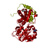
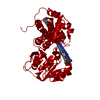
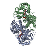
- PDB-1evy: CRYSTAL STRUCTURE OF LEISHMANIA MEXICANA GLYCEROL-3-PHOSPHATE DEH... -
+ Open data

- Basic information
| Entry | Database: PDB / ID: 1evy | ||||||
|---|---|---|---|---|---|---|---|
| Title | CRYSTAL STRUCTURE OF LEISHMANIA MEXICANA GLYCEROL-3-PHOSPHATE DEHYDROGENASE | ||||||
 Components Components | GLYCEROL-3-PHOSPHATE DEHYDROGENASE | ||||||
 Keywords Keywords | OXIDOREDUCTASE / Dehydrogenase / Rossmann fold | ||||||
| Function / homology |  Function and homology information Function and homology informationglycerol-3-phosphate dehydrogenase (NAD+) / glycerol-3-phosphate dehydrogenase (NAD+) activity / glycerol-3-phosphate catabolic process / glycosome / NAD binding / carbohydrate metabolic process / cytosol Similarity search - Function | ||||||
| Biological species |   Leishmania mexicana (eukaryote) Leishmania mexicana (eukaryote) | ||||||
| Method |  X-RAY DIFFRACTION / SYNCHROTRON / Resolution: 1.75 Å X-RAY DIFFRACTION / SYNCHROTRON / Resolution: 1.75 Å | ||||||
 Authors Authors | Suresh, S. / Turley, S. / Opperdoes, F.R. / Michels, P.A.M. / Hol, W.G.J. | ||||||
 Citation Citation | Journal: Structure Fold.Des. / Year: 2000 Title: A potential target enzyme for trypanocidal drugs revealed by the crystal structure of NAD-dependent glycerol-3-phosphate dehydrogenase from Leishmania mexicana. Authors: Suresh, S. / Turley, S. / Opperdoes, F.R. / Michels, P.A. / Hol, W.G. | ||||||
| History |
|
- Structure visualization
| Structure viewer | Molecule: MolmilJmol/JSmol |
|---|
- Downloads & links
-Download
| PDBx/mmCIF format | 1evy.cif.gz | 84.6 KB | Display | PDBx/mmCIF format |
|---|---|---|---|---|
| PDB format | pdb1evy.ent.gz | 64 KB | Display | PDB format |
| PDBx/mmJSON format | 1evy.json.gz | Tree view | PDBx/mmJSON format | |
| Others |  Other downloads Other downloads |
-Validation report
| Arichive directory | https://data.pdbj.org/pub/pdb/validation_reports/ev/1evyftp://data.pdbj.org/pub/pdb/validation_reports/ev/1evy | HTTPS FTP |
|---|
-Related structure data


-Links
 PDBj
PDBj


- Assembly
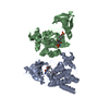
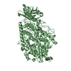
| Deposited unit | 
| ||||||||
|---|---|---|---|---|---|---|---|---|---|
| 1 | 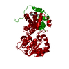
| ||||||||
| Unit cell |
| ||||||||
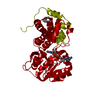
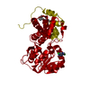
| Details | The biological assembly is a dimer constructed from chain A a symmetry partner generated by the two-fold. |
-Components
| #1: Protein | Mass: 39317.828 Da / Num. of mol.: 1 Source method: isolated from a genetically manipulated source Source: (gene. exp.) Leishmania mexicana (eukaryote) / Plasmid: PET3 / Production host:  References: UniProt: P90551, glycerol-3-phosphate dehydrogenase (NAD+) |
|---|---|
| #2: Chemical | ChemComp-MYS /   Mass: 212.415 Da / Num. of mol.: 1 / Source method: obtained synthetically / Formula: C15H32 Mass: 212.415 Da / Num. of mol.: 1 / Source method: obtained synthetically / Formula: C15H32 |
| #3: Water | ChemComp-HOH /  Mass: 18.015 Da / Num. of mol.: 328 / Source method: isolated from a natural source / Formula: H2O Mass: 18.015 Da / Num. of mol.: 328 / Source method: isolated from a natural source / Formula: H2O |
-Experimental details
-Experiment
| Experiment | Method: X-RAY DIFFRACTION / Number of used crystals: 1 |
|---|
- Sample preparation
| Crystal | Density Matthews: 3.3 Å3/Da / Density % sol: 62.75 % | ||||||||||||||||||||||||||||||||||||||||
|---|---|---|---|---|---|---|---|---|---|---|---|---|---|---|---|---|---|---|---|---|---|---|---|---|---|---|---|---|---|---|---|---|---|---|---|---|---|---|---|---|---|
| Crystal grow | Temperature: 298 K / Method: vapor diffusion, sitting drop / pH: 7.25 Details: Triethanol amine pH 7.25, Sodium Citrate 0.9 M, VAPOR DIFFUSION, SITTING DROP, temperature 298K | ||||||||||||||||||||||||||||||||||||||||
| Crystal grow | *PLUS pH: 7.2 | ||||||||||||||||||||||||||||||||||||||||
| Components of the solutions | *PLUS
|
-Data collection
| Diffraction | Mean temperature: 125 K |
|---|---|
| Diffraction source | Source: SYNCHROTRON / Site: SSRL  / Beamline: BL7-1 / Wavelength: 1.08 / Beamline: BL7-1 / Wavelength: 1.08 |
| Detector | Type: MARRESEARCH / Detector: IMAGE PLATE / Date: May 1, 1998 |
| Radiation | Protocol: SINGLE WAVELENGTH / Monochromatic (M) / Laue (L): M / Scattering type: x-ray |
| Radiation wavelength | Wavelength: 1.08 Å / Relative weight: 1 |
| Reflection | Resolution: 1.75→30 Å / Num. all: 46462 / Num. obs: 45712 / % possible obs: 85.4 % / Observed criterion σ(F): 2 / Observed criterion σ(I): 2 / Redundancy: 4.9 % / Biso Wilson estimate: 28.5 Å2 / Rmerge(I) obs: 0.044 / Net I/σ(I): 31.65 |
| Reflection shell | Resolution: 1.75→1.8 Å / Redundancy: 2 % / Rmerge(I) obs: 0.317 / Num. unique all: 1691 / % possible all: 63.7 |
| Reflection | *PLUS Num. measured all: 229249 |
| Reflection shell | *PLUS Lowest resolution: 1.8 Å / % possible obs: 63.7 % |
- Processing
| Software |
| ||||||||||||||||||||
|---|---|---|---|---|---|---|---|---|---|---|---|---|---|---|---|---|---|---|---|---|---|
| Refinement | Resolution: 1.75→30 Å / σ(F): 2 / σ(I): 2 / Stereochemistry target values: TNT
| ||||||||||||||||||||
| Refinement step | Cycle: LAST / Resolution: 1.75→30 Å
| ||||||||||||||||||||
| Refine LS restraints |
| ||||||||||||||||||||
| Software | *PLUS Name: TNT / Classification: refinement | ||||||||||||||||||||
| Refinement | *PLUS Lowest resolution: 30 Å / σ(F): 2 / % reflection Rfree: 5 % | ||||||||||||||||||||
| Solvent computation | *PLUS | ||||||||||||||||||||
| Displacement parameters | *PLUS | ||||||||||||||||||||
| Refine LS restraints | *PLUS Type: t_angle_deg / Dev ideal: 2.81 | ||||||||||||||||||||
| LS refinement shell | *PLUS Highest resolution: 1.75 Å / Lowest resolution: 1.8 Å / Rfactor Rfree: 0.38 / Rfactor obs: 0.36 |
