 Movie
Movie Controller

[English] 日本語
 Yorodumi
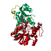
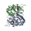
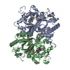
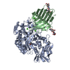
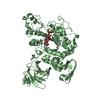
- PDB-4fzr: Crystal Structure of SsfS6, Streptomyces sp. SF2575 glycosyltrans... -
+ Open data

- Basic information
| Entry | Database: PDB / ID: 4fzr | ||||||
|---|---|---|---|---|---|---|---|
| Title | Crystal Structure of SsfS6, Streptomyces sp. SF2575 glycosyltransferase | ||||||
 Components Components | SsfS6 | ||||||
 Keywords Keywords | TRANSFERASE / Structural Genomics / PSI-Biology / Protein Structure Initiative / Enzyme Discovery for Natural Product Biosynthesis / NATPRO / Glycosyltransferase | ||||||
| Function / homology |  Function and homology information Function and homology informationUDP-glycosyltransferase activity / hexosyltransferase activity / antibiotic biosynthetic process Similarity search - Function | ||||||
| Biological species |  Streptomyces sp. SF2575 (bacteria) Streptomyces sp. SF2575 (bacteria) | ||||||
| Method |  X-RAY DIFFRACTION / SYNCHROTRON / SAD / Resolution: 2.397 Å X-RAY DIFFRACTION / SYNCHROTRON / SAD / Resolution: 2.397 Å | ||||||
 Authors Authors | Wang, F. / Zhou, M. / Singh, S. / Bingman, C.A. / Thorson, J.S. / Phillips Jr., G.N. / Enzyme Discovery for Natural Product Biosynthesis (NatPro) | ||||||
 Citation Citation | Journal: Proteins / Year: 2013 Title: Crystal structure of SsfS6, the putative C-glycosyltransferase involved in SF2575 biosynthesis. Authors: Wang, F. / Zhou, M. / Singh, S. / Yennamalli, R.M. / Bingman, C.A. / Thorson, J.S. / Phillips, G.N. | ||||||
| History |
|
- Structure visualization
| Structure viewer | Molecule: MolmilJmol/JSmol |
|---|
- Downloads & links
-Download
| PDBx/mmCIF format | 4fzr.cif.gz | 84.2 KB | Display | PDBx/mmCIF format |
|---|---|---|---|---|
| PDB format | pdb4fzr.ent.gz | 61.9 KB | Display | PDB format |
| PDBx/mmJSON format | 4fzr.json.gz | Tree view | PDBx/mmJSON format | |
| Others |  Other downloads Other downloads |
-Validation report
| Arichive directory | https://data.pdbj.org/pub/pdb/validation_reports/fz/4fzrftp://data.pdbj.org/pub/pdb/validation_reports/fz/4fzr | HTTPS FTP |
|---|
-Related structure data


-Links
 PDBj
PDBj- Assembly
| Deposited unit | 
| ||||||||
|---|---|---|---|---|---|---|---|---|---|
| 1 | 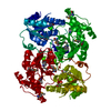
| ||||||||
| Unit cell |
|
-Components
| #1: Protein | Mass: 43225.000 Da / Num. of mol.: 1 Source method: isolated from a genetically manipulated source Source: (gene. exp.) Streptomyces sp. SF2575 (bacteria) / Gene: ssfS6 / Production host: |
|---|---|
| #2: Water | ChemComp-HOH /  Mass: 18.015 Da / Num. of mol.: 110 / Source method: isolated from a natural source / Formula: H2O Mass: 18.015 Da / Num. of mol.: 110 / Source method: isolated from a natural source / Formula: H2O |
| Has protein modification | Y |
-Experimental details
-Experiment
| Experiment | Method: X-RAY DIFFRACTION / Number of used crystals: 2 |
|---|
- Sample preparation
| Crystal | Density Matthews: 2.63 Å3/Da / Density % sol: 53.16 % |
|---|---|
| Crystal grow | Temperature: 298 K / Method: vapor diffusion, sitting drop / pH: 8.2 Details: Protein Solution (11.4 mg/ml SsfS6 protein, 50mM Tris pH 8.0) mixed in a 1:1 ratio with the well solution (1.13~1.40 M Sodium phosphate monobasic monohydrate / Potassium phosphate dibasic, ...Details: Protein Solution (11.4 mg/ml SsfS6 protein, 50mM Tris pH 8.0) mixed in a 1:1 ratio with the well solution (1.13~1.40 M Sodium phosphate monobasic monohydrate / Potassium phosphate dibasic, pH 8.2) Cryoprotected with 100% Paratone-N, VAPOR DIFFUSION, SITTING DROP, temperature 298K |
-Data collection
| Diffraction |
| ||||||||||||||||||||||||||||||||||||||||||||||||||||||||||||||||||||||||||||||||||||||||||||||||||||||||||||||||||||||||||||||
|---|---|---|---|---|---|---|---|---|---|---|---|---|---|---|---|---|---|---|---|---|---|---|---|---|---|---|---|---|---|---|---|---|---|---|---|---|---|---|---|---|---|---|---|---|---|---|---|---|---|---|---|---|---|---|---|---|---|---|---|---|---|---|---|---|---|---|---|---|---|---|---|---|---|---|---|---|---|---|---|---|---|---|---|---|---|---|---|---|---|---|---|---|---|---|---|---|---|---|---|---|---|---|---|---|---|---|---|---|---|---|---|---|---|---|---|---|---|---|---|---|---|---|---|---|---|---|---|
| Diffraction source |
| ||||||||||||||||||||||||||||||||||||||||||||||||||||||||||||||||||||||||||||||||||||||||||||||||||||||||||||||||||||||||||||||
| Detector |
| ||||||||||||||||||||||||||||||||||||||||||||||||||||||||||||||||||||||||||||||||||||||||||||||||||||||||||||||||||||||||||||||
| Radiation |
| ||||||||||||||||||||||||||||||||||||||||||||||||||||||||||||||||||||||||||||||||||||||||||||||||||||||||||||||||||||||||||||||
| Radiation wavelength |
| ||||||||||||||||||||||||||||||||||||||||||||||||||||||||||||||||||||||||||||||||||||||||||||||||||||||||||||||||||||||||||||||
| Reflection | Resolution: 2.4→50 Å / Num. all: 19209 / Num. obs: 19017 / % possible obs: 99 % / Observed criterion σ(F): 0 / Observed criterion σ(I): 0 / Redundancy: 11.5 % / Biso Wilson estimate: 70.76 Å2 / Rmerge(I) obs: 0.094 / Χ2: 0.923 / Net I/σ(I): 8.7 | ||||||||||||||||||||||||||||||||||||||||||||||||||||||||||||||||||||||||||||||||||||||||||||||||||||||||||||||||||||||||||||||
| Reflection shell |
|

- Processing
| Software |
| ||||||||||||||||||||||||||||||||||||||||||||||||||||||||
|---|---|---|---|---|---|---|---|---|---|---|---|---|---|---|---|---|---|---|---|---|---|---|---|---|---|---|---|---|---|---|---|---|---|---|---|---|---|---|---|---|---|---|---|---|---|---|---|---|---|---|---|---|---|---|---|---|---|
| Refinement | Method to determine structure: SAD / Resolution: 2.397→30.763 Å / Occupancy max: 1 / Occupancy min: 0.48 / FOM work R set: 0.7525 / SU ML: 0.32 / σ(F): 1.34 / Phase error: 29.8 / Stereochemistry target values: ML
| ||||||||||||||||||||||||||||||||||||||||||||||||||||||||
| Solvent computation | Shrinkage radii: 0.9 Å / VDW probe radii: 1.11 Å / Solvent model: FLAT BULK SOLVENT MODEL | ||||||||||||||||||||||||||||||||||||||||||||||||||||||||
| Displacement parameters | Biso max: 133.15 Å2 / Biso mean: 39.7201 Å2 / Biso min: 6.7 Å2 | ||||||||||||||||||||||||||||||||||||||||||||||||||||||||
| Refinement step | Cycle: LAST / Resolution: 2.397→30.763 Å
| ||||||||||||||||||||||||||||||||||||||||||||||||||||||||
| Refine LS restraints |
| ||||||||||||||||||||||||||||||||||||||||||||||||||||||||
| LS refinement shell | Refine-ID: X-RAY DIFFRACTION / Total num. of bins used: 7
|
