- PDB-6gzw: Ferric DtpA from Streptomyces lividans -
+
Open data
ID or keywords:
Loading...
-
Basic information
Entry
Database: PDB / ID: 6gzw
Title
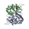
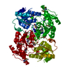
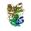
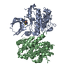
Ferric DtpA from Streptomyces lividans
Components
Dye type peroxidase A
Keywords
OXIDOREDUCTASE / peroxidase / ferric / dye-type
Function / homology
Function and homology information
iron import into cell / Oxidoreductases; Acting on a peroxide as acceptor; Peroxidases / peroxidase activity / heme binding / metal ion binding Similarity search - Function
Mass: 18.015 Da / Num. of mol.: 793 / Source method: isolated from a natural source / Formula: H2O
-
Experimental details
-
Experiment
Experiment
Method: X-RAY DIFFRACTION / Number of used crystals: 1
-
Sample preparation
Crystal
Density Matthews: 2.09 Å3/Da / Density % sol: 41.01 %
Crystal grow
Temperature: 291 K / Method: vapor diffusion, hanging drop / pH: 5.5 Details: Crystals were grown by the vapour diffusion hanging drop method using a ferric protein solution at 13 mg/ml equilibrated against a reservoir consisting of 17-24% PEG 3000 and 50-100 mM sodium citrate, pH 5.5.
Resolution: 1.41→77.99 Å / Cor.coef. Fo:Fc: 0.965 / Cor.coef. Fo:Fc free: 0.955 / SU B: 1.757 / SU ML: 0.064 / SU R Cruickshank DPI: 0.0768 / Cross valid method: THROUGHOUT / σ(F): 0 / ESU R: 0.077 / ESU R Free: 0.077 Details: HYDROGENS HAVE BEEN ADDED IN THE RIDING POSITIONS U VALUES : REFINED INDIVIDUALLY
Rfactor
Num. reflection
% reflection
Selection details
Rfree
0.2163
5945
5 %
RANDOM
Rwork
0.1891
-
-
-
obs
0.1905
112316
93.63 %
-
Solvent computation
Ion probe radii: 0.8 Å / Shrinkage radii: 0.8 Å / VDW probe radii: 1.2 Å
In the structure databanks used in Yorodumi, some data are registered as the other names, "COVID-19 virus" and "2019-nCoV". Here are the details of the virus and the list of structure data.
Jan 31, 2019. EMDB accession codes are about to change! (news from PDBe EMDB page)
EMDB accession codes are about to change! (news from PDBe EMDB page)
The allocation of 4 digits for EMDB accession codes will soon come to an end. Whilst these codes will remain in use, new EMDB accession codes will include an additional digit and will expand incrementally as the available range of codes is exhausted. The current 4-digit format prefixed with “EMD-” (i.e. EMD-XXXX) will advance to a 5-digit format (i.e. EMD-XXXXX), and so on. It is currently estimated that the 4-digit codes will be depleted around Spring 2019, at which point the 5-digit format will come into force.
The EM Navigator/Yorodumi systems omit the EMD- prefix.
Related info.:Q: What is EMD? / ID/Accession-code notation in Yorodumi/EM Navigator
Yorodumi is a browser for structure data from EMDB, PDB, SASBDB, etc.
This page is also the successor to EM Navigator detail page, and also detail information page/front-end page for Omokage search.
The word "yorodu" (or yorozu) is an old Japanese word meaning "ten thousand". "mi" (miru) is to see.
Related info.:EMDB / PDB / SASBDB / Comparison of 3 databanks / Yorodumi Search / Aug 31, 2016. New EM Navigator & Yorodumi / Yorodumi Papers / Jmol/JSmol / Function and homology information / Changes in new EM Navigator and Yorodumi
 Movie
Movie Controller


 Open data
 Basic information
 Components
 Keywords
 Function and homology information
 Streptomyces lividans 1326 (bacteria)
 X-RAY DIFFRACTION /
  Authors
 United Kingdom, 1items
  Citation
 Structure visualization
 Downloads & links
 Other downloads


 PDBj
PDBj

 Assembly

 Mass: 616.487 Da / Num. of mol.: 2 / Source method: obtained synthetically / Formula: C34H32FeN4O4
 Mass: 18.015 Da / Num. of mol.: 793 / Source method: isolated from a natural source / Formula: H2O
 Sample preparation
 / Beamline: MASSIF-3 / Wavelength: 0.9677 Å
 Processing