 Movie
Movie Controller

[English] 日本語
 Yorodumi
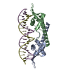
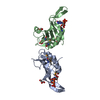
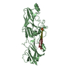
- PDB-1j0r: Crystal structure of the replication termination protein mutant C110S -
+ Open data

- Basic information
| Entry | Database: PDB / ID: 1j0r | ||||||
|---|---|---|---|---|---|---|---|
| Title | Crystal structure of the replication termination protein mutant C110S | ||||||
 Components Components | replication termination protein | ||||||
 Keywords Keywords | REPLICATION / winged-helix / DNA-binding protein | ||||||
| Function / homology |  Function and homology information Function and homology information | ||||||
| Biological species |  | ||||||
| Method |  X-RAY DIFFRACTION / MOLECULAR REPLACEMENT / Resolution: 2.5 Å X-RAY DIFFRACTION / MOLECULAR REPLACEMENT / Resolution: 2.5 Å | ||||||
 Authors Authors | Vivian, J.P. / Hastings, A.F. / Duggin, I.G. / Wake, R.G. / Wilce, M.C.J. / Wilce, J.A. | ||||||
 Citation Citation | Journal: Biochem.Biophys.Res.Commun. / Year: 2003 Title: The impact of single cysteine residue mutations on the replication terminator protein Authors: Vivian, J.P. / Hastings, A.F. / Duggin, I.G. / Wake, R.G. / Wilce, M.C.J. / Wilce, J.A. | ||||||
| History |
|
- Structure visualization
| Structure viewer | Molecule: MolmilJmol/JSmol |
|---|
- Downloads & links
-Download
| PDBx/mmCIF format | 1j0r.cif.gz | 59.3 KB | Display | PDBx/mmCIF format |
|---|---|---|---|---|
| PDB format | pdb1j0r.ent.gz | 44.1 KB | Display | PDB format |
| PDBx/mmJSON format | 1j0r.json.gz | Tree view | PDBx/mmJSON format | |
| Others |  Other downloads Other downloads |
-Validation report
| Arichive directory | https://data.pdbj.org/pub/pdb/validation_reports/j0/1j0rftp://data.pdbj.org/pub/pdb/validation_reports/j0/1j0r | HTTPS FTP |
|---|
-Related structure data
| Related structure data | 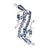 1bm9S S: Starting model for refinement |
|---|---|
| Similar structure data |


-Links
 PDBj
PDBj- Assembly
| Deposited unit | 
| ||||||||
|---|---|---|---|---|---|---|---|---|---|
| 1 | 
| ||||||||
| 2 | 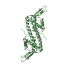
| ||||||||
| Unit cell |
|
-Components
| #1: Protein | Mass: 14531.099 Da / Num. of mol.: 2 / Mutation: C110S Source method: isolated from a genetically manipulated source Source: (gene. exp.) #2: Water | ChemComp-HOH / |  Mass: 18.015 Da / Num. of mol.: 58 / Source method: isolated from a natural source / Formula: H2O Mass: 18.015 Da / Num. of mol.: 58 / Source method: isolated from a natural source / Formula: H2O |
|---|
-Experimental details
-Experiment
| Experiment | Method: X-RAY DIFFRACTION / Number of used crystals: 1 |
|---|
- Sample preparation
| Crystal | Density Matthews: 2.45 Å3/Da / Density % sol: 49.32 % | ||||||||||||||||||||||||||||||
|---|---|---|---|---|---|---|---|---|---|---|---|---|---|---|---|---|---|---|---|---|---|---|---|---|---|---|---|---|---|---|---|
| Crystal grow | Temperature: 295 K / Method: vapor diffusion, hanging drop / pH: 6 Details: 22% PEG 2000, 150mM MES, pH 6.0, VAPOR DIFFUSION, HANGING DROP, temperature 295K | ||||||||||||||||||||||||||||||
| Crystal grow | *PLUS Temperature: 22 ℃ / pH: 6.7 / Method: vapor diffusion, hanging drop | ||||||||||||||||||||||||||||||
| Components of the solutions | *PLUS
|
-Data collection
| Diffraction | Mean temperature: 100 K |
|---|---|
| Diffraction source | Source: ROTATING ANODE / Type: RIGAKU RU200 / Wavelength: 1.54 Å |
| Detector | Type: MARRESEARCH / Detector: IMAGE PLATE / Date: Jun 7, 2001 / Details: mirrors |
| Radiation | Monochromator: mirrors / Protocol: SINGLE WAVELENGTH / Monochromatic (M) / Laue (L): M / Scattering type: x-ray |
| Radiation wavelength | Wavelength: 1.54 Å / Relative weight: 1 |
| Reflection | Resolution: 2.5→69 Å / Num. all: 9541 / Num. obs: 9541 / % possible obs: 88 % / Observed criterion σ(I): 2 / Redundancy: 5.7 % / Rsym value: 0.045 |
| Reflection shell | Resolution: 2.5→2.59 Å / Rsym value: 0.471 / % possible all: 92.2 |
| Reflection | *PLUS % possible obs: 88 % / Num. measured all: 54110 / Rmerge(I) obs: 0.045 |
| Reflection shell | *PLUS Highest resolution: 2.5 Å / Lowest resolution: 2.66 Å / % possible obs: 92.2 % / Rmerge(I) obs: 0.471 |
- Processing
| Software |
| ||||||||||||||||||||
|---|---|---|---|---|---|---|---|---|---|---|---|---|---|---|---|---|---|---|---|---|---|
| Refinement | Method to determine structure: MOLECULAR REPLACEMENT Starting model: PDB entry 1BM9 Resolution: 2.5→50 Å / σ(F): 0 / Stereochemistry target values: Engh & Huber
| ||||||||||||||||||||
| Refinement step | Cycle: LAST / Resolution: 2.5→50 Å
| ||||||||||||||||||||
| Refine LS restraints |
| ||||||||||||||||||||
| LS refinement shell | Resolution: 2.5→2.59 Å
| ||||||||||||||||||||
| Refine LS restraints | *PLUS
| ||||||||||||||||||||
| LS refinement shell | *PLUS Highest resolution: 2.5 Å / Lowest resolution: 2.66 Å |
