 Movie
Movie Controller

[English] 日本語
 Yorodumi
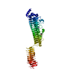
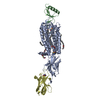
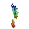
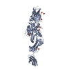
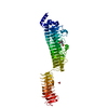
- PDB-4nuv: Heterotetramer structure of Region II from Plasmodium vivax Duffy... -
+ Open data

- Basic information
| Entry | Database: PDB / ID: 4nuv | ||||||
|---|---|---|---|---|---|---|---|
| Title | Heterotetramer structure of Region II from Plasmodium vivax Duffy Binding Protein (PvDBP) bound to the ectodomain of the Duffy Antigen Receptor for Chemokines (DARC) | ||||||
 Components Components |
| ||||||
 Keywords Keywords | membrane protein / cell invasion / Duffy Binding Like (DBL) Domain Fold / GPCR / Adhesion / Invasion / Red blood cell binding / Chemokine Binding / Duffy Antigen Receptor for Chemokines / Membrane / protein binding | ||||||
| Function / homology |  Function and homology information Function and homology informationregulation of chemokine production / C-C chemokine binding / Peptide ligand-binding receptors / chemokine-mediated signaling pathway / defense response / recycling endosome / G protein-coupled receptor activity / transmembrane signaling receptor activity / signaling receptor activity / early endosome ...regulation of chemokine production / C-C chemokine binding / Peptide ligand-binding receptors / chemokine-mediated signaling pathway / defense response / recycling endosome / G protein-coupled receptor activity / transmembrane signaling receptor activity / signaling receptor activity / early endosome / host cell surface receptor binding / inflammatory response / membrane / identical protein binding / plasma membrane Similarity search - Function | ||||||
| Biological species |   Homo sapiens (human) Homo sapiens (human) | ||||||
| Method |  X-RAY DIFFRACTION / SYNCHROTRON / MOLECULAR REPLACEMENT / Resolution: 2.6 Å X-RAY DIFFRACTION / SYNCHROTRON / MOLECULAR REPLACEMENT / Resolution: 2.6 Å | ||||||
 Authors Authors | Tolia, N.H. | ||||||
 Citation Citation | Journal: Plos Pathog. / Year: 2014 Title: Red Blood Cell Invasion by Plasmodium vivax: Structural Basis for DBP Engagement of DARC. Authors: Batchelor, J.D. / Malpede, B.M. / Omattage, N.S. / Dekoster, G.T. / Henzler-Wildman, K.A. / Tolia, N.H. | ||||||
| History |
|
- Structure visualization
| Structure viewer | Molecule: MolmilJmol/JSmol |
|---|
- Downloads & links
-Download
| PDBx/mmCIF format | 4nuv.cif.gz | 376.6 KB | Display | PDBx/mmCIF format |
|---|---|---|---|---|
| PDB format | pdb4nuv.ent.gz | 316.6 KB | Display | PDB format |
| PDBx/mmJSON format | 4nuv.json.gz | Tree view | PDBx/mmJSON format | |
| Others |  Other downloads Other downloads |
-Validation report
| Arichive directory | https://data.pdbj.org/pub/pdb/validation_reports/nu/4nuvftp://data.pdbj.org/pub/pdb/validation_reports/nu/4nuv | HTTPS FTP |
|---|
-Related structure data
| Related structure data |  4nuuC  3rrcS C: citing same article ( S: Starting model for refinement |
|---|---|
| Similar structure data |


-Links
 PDBj
PDBj


- Assembly
| Deposited unit | 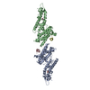
| ||||||||
|---|---|---|---|---|---|---|---|---|---|
| 1 |
| ||||||||
| 2 | 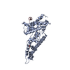
| ||||||||
| 3 | 
| ||||||||
| Unit cell |
|
-Components
| #1: Protein | Mass: 37610.051 Da / Num. of mol.: 2 / Fragment: unp residues 211-525 Source method: isolated from a genetically manipulated source Source: (gene. exp.) Strain: Salvador I / Gene: PVDR / Production host:  #2: Protein/peptide | Mass: 3628.606 Da / Num. of mol.: 2 / Fragment: unp residues 14-43 Source method: isolated from a genetically manipulated source Source: (gene. exp.) Homo sapiens (human) / Gene: DARC, ACKR1, FY, GPD / Production host: #3: Chemical |   Mass: 92.094 Da / Num. of mol.: 2 / Source method: obtained synthetically / Formula: C3H8O3 Mass: 92.094 Da / Num. of mol.: 2 / Source method: obtained synthetically / Formula: C3H8O3#4: Water | ChemComp-HOH / |  Mass: 18.015 Da / Num. of mol.: 52 / Source method: isolated from a natural source / Formula: H2O Mass: 18.015 Da / Num. of mol.: 52 / Source method: isolated from a natural source / Formula: H2OHas protein modification | Y | |
|---|
-Experimental details
-Experiment
| Experiment | Method: X-RAY DIFFRACTION / Number of used crystals: 1 |
|---|
- Sample preparation
| Crystal | Density Matthews: 2.35 Å3/Da / Density % sol: 47.59 % |
|---|---|
| Crystal grow | Temperature: 290 K / Method: vapor diffusion, hanging drop / pH: 7.4 Details: 0.1 M HEPES and 20% (w/v) polyethylene glycol 6000, VAPOR DIFFUSION, HANGING DROP, temperature 290K, pH 7.4 |
-Data collection
| Diffraction | Mean temperature: 93 K |
|---|---|
| Diffraction source | Source: SYNCHROTRON / Site: ALS  / Beamline: 4.2.2 / Wavelength: 1 Å / Beamline: 4.2.2 / Wavelength: 1 Å |
| Detector | Type: NOIR-1 / Detector: CCD / Date: Dec 20, 2011 |
| Radiation | Monochromator: Rosenbaum-Rock monochromator 1: high-resolution double-crystal sagittal focusing, Rosenbaum-Rock monochromator 2: double crystal, Rosenbaum-Rock vertical focusing mirror Protocol: SINGLE WAVELENGTH / Monochromatic (M) / Laue (L): M / Scattering type: x-ray |
| Radiation wavelength | Wavelength: 1 Å / Relative weight: 1 |
| Reflection | Resolution: 2.6→20 Å / Num. all: 23251 / Num. obs: 22139 / % possible obs: 95.2 % / Observed criterion σ(F): 0 / Observed criterion σ(I): 0 |
| Reflection shell | Resolution: 2.6→2.7 Å / % possible all: 93.1 |
- Processing
| Software |
| |||||||||||||||||||||||||||||||||||||||||||||||||||||||||||||||
|---|---|---|---|---|---|---|---|---|---|---|---|---|---|---|---|---|---|---|---|---|---|---|---|---|---|---|---|---|---|---|---|---|---|---|---|---|---|---|---|---|---|---|---|---|---|---|---|---|---|---|---|---|---|---|---|---|---|---|---|---|---|---|---|---|
| Refinement | Method to determine structure: MOLECULAR REPLACEMENT Starting model: pdb entry 3RRC Resolution: 2.6→19.904 Å / SU ML: 0.32 / σ(F): 1.99 / Phase error: 25.69 / Stereochemistry target values: ML
| |||||||||||||||||||||||||||||||||||||||||||||||||||||||||||||||
| Solvent computation | Shrinkage radii: 0.9 Å / VDW probe radii: 1.11 Å / Solvent model: FLAT BULK SOLVENT MODEL | |||||||||||||||||||||||||||||||||||||||||||||||||||||||||||||||
| Refinement step | Cycle: LAST / Resolution: 2.6→19.904 Å
| |||||||||||||||||||||||||||||||||||||||||||||||||||||||||||||||
| Refine LS restraints |
| |||||||||||||||||||||||||||||||||||||||||||||||||||||||||||||||
| LS refinement shell |
|
