 Movie
Movie Controller

[English] 日本語
 Yorodumi
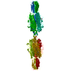
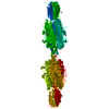
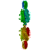
- PDB-3gqa: Crystal Structure of the Bacteriophage phi29 gene product 12 N-te... -
+ Open data

- Basic information
| Entry | Database: PDB / ID: 3gqa | ||||||
|---|---|---|---|---|---|---|---|
| Title | Crystal Structure of the Bacteriophage phi29 gene product 12 N-terminal fragment in complex with cobalt ions | ||||||
 Components Components | Preneck appendage protein | ||||||
 Keywords Keywords | VIRAL PROTEIN / beta helix | ||||||
| Function / homology |  Function and homology information Function and homology informationvirus tail, fiber / symbiont entry into host cell via disruption of host cell envelope / adhesion receptor-mediated virion attachment to host cell / virion attachment to host cell / ATP binding / metal ion binding Similarity search - Function | ||||||
| Biological species |   Bacillus phage phi29 (virus) Bacillus phage phi29 (virus) | ||||||
| Method |  X-RAY DIFFRACTION / SYNCHROTRON / MOLECULAR REPLACEMENT / Resolution: 2.1 Å X-RAY DIFFRACTION / SYNCHROTRON / MOLECULAR REPLACEMENT / Resolution: 2.1 Å | ||||||
 Authors Authors | Xiang, Y. / Rossmann, M.G. | ||||||
 Citation Citation | Journal: Mol.Cell / Year: 2009 Title: Crystallographic insights into the autocatalytic assembly mechanism of a bacteriophage tail spike. Authors: Xiang, Y. / Leiman, P.G. / Li, L. / Grimes, S. / Anderson, D.L. / Rossmann, M.G. | ||||||
| History |
|
- Structure visualization
| Structure viewer | Molecule: MolmilJmol/JSmol |
|---|
- Downloads & links
-Download
| PDBx/mmCIF format | 3gqa.cif.gz | 130.6 KB | Display | PDBx/mmCIF format |
|---|---|---|---|---|
| PDB format | pdb3gqa.ent.gz | 99.7 KB | Display | PDB format |
| PDBx/mmJSON format | 3gqa.json.gz | Tree view | PDBx/mmJSON format | |
| Others |  Other downloads Other downloads |
-Validation report
| Arichive directory | https://data.pdbj.org/pub/pdb/validation_reports/gq/3gqaftp://data.pdbj.org/pub/pdb/validation_reports/gq/3gqa | HTTPS FTP |
|---|
-Related structure data
| Related structure data | 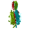 3gq7SC 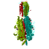 3gq8C  3gq9C 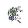 3gqhC  3gqkC 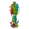 3sucC S: Starting model for refinement C: citing same article ( |
|---|---|
| Similar structure data |


-Links
 PDBj
PDBj
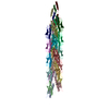
- Assembly
| Deposited unit | 
| ||||||||
|---|---|---|---|---|---|---|---|---|---|
| 1 | x 6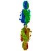
| ||||||||
| 2 | 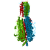
| ||||||||
| Unit cell |
| ||||||||
| Components on special symmetry positions |
|
-Components
| #1: Protein | Mass: 64410.914 Da / Num. of mol.: 1 / Fragment: D1*D2D3, Residues 89-691 Source method: isolated from a genetically manipulated source Details: RESIDUES 89-854 EXPRESSED WITH A N-TERMIINAL HIS6 TAG Source: (gene. exp.) Bacillus phage phi29 (virus) / Gene: 12, gene product 12 / Plasmid: PET28B / Production host:  | ||||||
|---|---|---|---|---|---|---|---|
| #2: Chemical |   Mass: 58.933 Da / Num. of mol.: 2 / Source method: obtained synthetically / Formula: Co Mass: 58.933 Da / Num. of mol.: 2 / Source method: obtained synthetically / Formula: Co#3: Chemical | ChemComp-PO4 / |   Mass: 94.971 Da / Num. of mol.: 1 / Source method: obtained synthetically / Formula: PO4 Mass: 94.971 Da / Num. of mol.: 1 / Source method: obtained synthetically / Formula: PO4#4: Water | ChemComp-HOH / |  Mass: 18.015 Da / Num. of mol.: 220 / Source method: isolated from a natural source / Formula: H2O Mass: 18.015 Da / Num. of mol.: 220 / Source method: isolated from a natural source / Formula: H2OSequence details | THESE MUTATIONS MIGHT BE AN ACCUMULATE | |
-Experimental details
-Experiment
| Experiment | Method: X-RAY DIFFRACTION / Number of used crystals: 1 |
|---|
- Sample preparation
| Crystal | Density Matthews: 3.52 Å3/Da / Density % sol: 65.04 % |
|---|---|
| Crystal grow | Temperature: 298 K / Method: vapor diffusion, hanging drop / pH: 10.5 Details: 100mM Tris-base at pH 10.5 and 8% PEG3K. Apo crystals were obtained by soaking the crystals in a mother liquid containing 10mM EDTA. The cobalt derivative was obtained by back soaking the ...Details: 100mM Tris-base at pH 10.5 and 8% PEG3K. Apo crystals were obtained by soaking the crystals in a mother liquid containing 10mM EDTA. The cobalt derivative was obtained by back soaking the apo-crystals in a mother liquid containing 10mM cobalt ions, VAPOR DIFFUSION, HANGING DROP, temperature 298K |
-Data collection
| Diffraction | Mean temperature: 100 K | |||||||||||||||||||||||||||||||||||||||||||||||||||||||||||||||||||||||||||||
|---|---|---|---|---|---|---|---|---|---|---|---|---|---|---|---|---|---|---|---|---|---|---|---|---|---|---|---|---|---|---|---|---|---|---|---|---|---|---|---|---|---|---|---|---|---|---|---|---|---|---|---|---|---|---|---|---|---|---|---|---|---|---|---|---|---|---|---|---|---|---|---|---|---|---|---|---|---|---|
| Diffraction source | Source: SYNCHROTRON / Site: APS  / Beamline: 23-ID-D / Wavelength: 0.98 Å / Beamline: 23-ID-D / Wavelength: 0.98 Å | |||||||||||||||||||||||||||||||||||||||||||||||||||||||||||||||||||||||||||||
| Detector | Type: MARMOSAIC 300 mm CCD / Detector: CCD / Date: Apr 21, 2008 | |||||||||||||||||||||||||||||||||||||||||||||||||||||||||||||||||||||||||||||
| Radiation | Monochromator: SI(111) double-crystal / Protocol: SINGLE WAVELENGTH / Monochromatic (M) / Laue (L): M / Scattering type: x-ray | |||||||||||||||||||||||||||||||||||||||||||||||||||||||||||||||||||||||||||||
| Radiation wavelength | Wavelength: 0.98 Å / Relative weight: 1 | |||||||||||||||||||||||||||||||||||||||||||||||||||||||||||||||||||||||||||||
| Reflection | Resolution: 2.1→43.88 Å / Num. obs: 53528 / % possible obs: 99.5 % / Redundancy: 8.5 % / Rmerge(I) obs: 0.099 / Χ2: 1.843 / Net I/σ(I): 31.667 | |||||||||||||||||||||||||||||||||||||||||||||||||||||||||||||||||||||||||||||
| Reflection shell |
|
- Processing
| Software |
| ||||||||||||||||||||||||||||||||||||||||||||||||||||||||||||||||||||||||||||||||||||||||||
|---|---|---|---|---|---|---|---|---|---|---|---|---|---|---|---|---|---|---|---|---|---|---|---|---|---|---|---|---|---|---|---|---|---|---|---|---|---|---|---|---|---|---|---|---|---|---|---|---|---|---|---|---|---|---|---|---|---|---|---|---|---|---|---|---|---|---|---|---|---|---|---|---|---|---|---|---|---|---|---|---|---|---|---|---|---|---|---|---|---|---|---|
| Refinement | Method to determine structure: MOLECULAR REPLACEMENT Starting model: 3GQ7 Resolution: 2.1→43.88 Å / Cor.coef. Fo:Fc: 0.963 / Cor.coef. Fo:Fc free: 0.942 / Occupancy max: 1 / Occupancy min: 0.33 / SU B: 3.317 / SU ML: 0.09 / Cross valid method: THROUGHOUT / σ(F): 0 / ESU R: 0.148 / ESU R Free: 0.145 / Stereochemistry target values: MAXIMUM LIKELIHOOD
| ||||||||||||||||||||||||||||||||||||||||||||||||||||||||||||||||||||||||||||||||||||||||||
| Solvent computation | Ion probe radii: 0.8 Å / Shrinkage radii: 0.8 Å / VDW probe radii: 1.2 Å / Solvent model: BABINET MODEL WITH MASK | ||||||||||||||||||||||||||||||||||||||||||||||||||||||||||||||||||||||||||||||||||||||||||
| Displacement parameters | Biso max: 74.63 Å2 / Biso mean: 29.128 Å2 / Biso min: 16.21 Å2
| ||||||||||||||||||||||||||||||||||||||||||||||||||||||||||||||||||||||||||||||||||||||||||
| Refinement step | Cycle: LAST / Resolution: 2.1→43.88 Å
| ||||||||||||||||||||||||||||||||||||||||||||||||||||||||||||||||||||||||||||||||||||||||||
| Refine LS restraints |
| ||||||||||||||||||||||||||||||||||||||||||||||||||||||||||||||||||||||||||||||||||||||||||
| LS refinement shell | Resolution: 2.1→2.157 Å / Total num. of bins used: 20
|
