 Movie
Movie Controller

[English] 日本語
 Yorodumi
- PDB-3suc: Crystal structure of the pre-mature bacteriophage phi29 gene prod... -
+ Open data

- Basic information
| Entry | Database: PDB / ID: 3suc | |||||||||
|---|---|---|---|---|---|---|---|---|---|---|
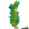
| Title | Crystal structure of the pre-mature bacteriophage phi29 gene product 12 | |||||||||
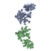
 Components Components | Preneck appendage protein | |||||||||
 Keywords Keywords | VIRAL PROTEIN / BETA HELIX / BETA BARREL / ATP BINDING | |||||||||
| Function / homology |  Function and homology information Function and homology informationvirus tail, fiber / symbiont entry into host cell via disruption of host cell envelope / adhesion receptor-mediated virion attachment to host cell / virion attachment to host cell / ATP binding / metal ion binding Similarity search - Function | |||||||||
| Biological species |   Bacillus phage phi29 (virus) Bacillus phage phi29 (virus) | |||||||||
| Method |  X-RAY DIFFRACTION / SYNCHROTRON / MOLECULAR REPLACEMENT / Resolution: 2.15 Å X-RAY DIFFRACTION / SYNCHROTRON / MOLECULAR REPLACEMENT / Resolution: 2.15 Å | |||||||||
 Authors Authors | Xiang, Y. / Rossmann, M.G. | |||||||||
 Citation Citation | Journal: Mol.Cell / Year: 2009 Title: Crystallographic Insights Into the Autocatalytic Assembly Mechanism of a Bacteriophage Tail Spike Authors: Xiang, Y. / G Leiman, P. / Li, L. / Grimes, S. / Anderson, D.L. / Rossmann, M.G. | |||||||||
| History |
|
- Structure visualization
| Structure viewer | Molecule: MolmilJmol/JSmol |
|---|
- Downloads & links
-Download
| PDBx/mmCIF format | 3suc.cif.gz | 305.8 KB | Display | PDBx/mmCIF format |
|---|---|---|---|---|
| PDB format | pdb3suc.ent.gz | 247.3 KB | Display | PDB format |
| PDBx/mmJSON format | 3suc.json.gz | Tree view | PDBx/mmJSON format | |
| Others |  Other downloads Other downloads |
-Validation report
| Arichive directory | https://data.pdbj.org/pub/pdb/validation_reports/su/3sucftp://data.pdbj.org/pub/pdb/validation_reports/su/3suc | HTTPS FTP |
|---|
-Related structure data
| Related structure data | 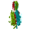 3gq7SC 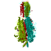 3gq8C  3gq9C 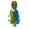 3gqaC  3gqhSC  3gqkC S: Starting model for refinement C: citing same article ( |
|---|---|
| Similar structure data |


-Links
 PDBj
PDBj- Assembly
| Deposited unit | 
| ||||||||
|---|---|---|---|---|---|---|---|---|---|
| 1 | 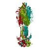
| ||||||||
| 2 | x 6
| ||||||||
| Unit cell |
|
-Components
| #1: Protein | Mass: 82406.320 Da / Num. of mol.: 1 / Fragment: D1*D2D3D4, RESIDUES 89-854 / Mutation: K166R, K627Q, E695Q, D701G, G817S Source method: isolated from a genetically manipulated source Source: (gene. exp.) Bacillus phage phi29 (virus) / Gene: 12 / Plasmid: PET28B / Production host:  | ||||
|---|---|---|---|---|---|
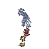
| #2: Chemical | ChemComp-CA /   Mass: 40.078 Da / Num. of mol.: 1 / Source method: obtained synthetically / Formula: Ca Mass: 40.078 Da / Num. of mol.: 1 / Source method: obtained synthetically / Formula: Ca | ||||
| #3: Chemical |   Mass: 24.305 Da / Num. of mol.: 2 / Source method: obtained synthetically / Formula: Mg Mass: 24.305 Da / Num. of mol.: 2 / Source method: obtained synthetically / Formula: Mg#4: Chemical | ChemComp-ATP / |   Mass: 507.181 Da / Num. of mol.: 1 / Source method: obtained synthetically / Formula: C10H16N5O13P3 / Comment: ATP, energy-carrying molecule*YM Mass: 507.181 Da / Num. of mol.: 1 / Source method: obtained synthetically / Formula: C10H16N5O13P3 / Comment: ATP, energy-carrying molecule*YM#5: Water | ChemComp-HOH / |  Mass: 18.015 Da / Num. of mol.: 73 / Source method: isolated from a natural source / Formula: H2O Mass: 18.015 Da / Num. of mol.: 73 / Source method: isolated from a natural source / Formula: H2O |
-Experimental details
-Experiment
| Experiment | Method: X-RAY DIFFRACTION / Number of used crystals: 1 |
|---|
- Sample preparation
| Crystal | Density Matthews: 4.27 Å3/Da / Density % sol: 71.19 % |
|---|---|
| Crystal grow | Temperature: 298 K / Method: vapor diffusion, hanging drop / pH: 7 Details: 3.0 M Sodium Acetate at pH 7.0, VAPOR DIFFUSION, HANGING DROP, temperature 298K |
-Data collection
| Diffraction | Mean temperature: 100 K |
|---|---|
| Diffraction source | Source: SYNCHROTRON / Site: APS  / Beamline: 23-ID-D / Wavelength: 0.98 / Beamline: 23-ID-D / Wavelength: 0.98 |
| Detector | Type: MARMOSAIC 300 mm CCD / Detector: CCD / Date: Jun 28, 2008 |
| Radiation | Monochromator: SI(111) DOUBLE-CRYSTAL / Protocol: SINGLE WAVELENGTH / Monochromatic (M) / Laue (L): M / Scattering type: x-ray |
| Radiation wavelength | Wavelength: 0.98 Å / Relative weight: 1 |
| Reflection | Resolution: 2.15→49.27 Å / Num. obs: 68349 / % possible obs: 88 % / Redundancy: 15.6 % / Rmerge(I) obs: 0.146 / Net I/σ(I): 17.143 |
| Reflection shell | Resolution: 2.15→2.23 Å / Redundancy: 6 % / Rmerge(I) obs: 0.446 / % possible all: 57 |
- Processing
| Software |
| ||||||||||||||||||||||||||||||||||||||||||||||||||||||||||||||||||||||||||||||||||||||||||||||||||||||||||||||||||||||||||||||||||||||||||||||||||||||||||||||||||||||||||
|---|---|---|---|---|---|---|---|---|---|---|---|---|---|---|---|---|---|---|---|---|---|---|---|---|---|---|---|---|---|---|---|---|---|---|---|---|---|---|---|---|---|---|---|---|---|---|---|---|---|---|---|---|---|---|---|---|---|---|---|---|---|---|---|---|---|---|---|---|---|---|---|---|---|---|---|---|---|---|---|---|---|---|---|---|---|---|---|---|---|---|---|---|---|---|---|---|---|---|---|---|---|---|---|---|---|---|---|---|---|---|---|---|---|---|---|---|---|---|---|---|---|---|---|---|---|---|---|---|---|---|---|---|---|---|---|---|---|---|---|---|---|---|---|---|---|---|---|---|---|---|---|---|---|---|---|---|---|---|---|---|---|---|---|---|---|---|---|---|---|---|---|
| Refinement | Method to determine structure: MOLECULAR REPLACEMENT Starting model: PDB entries 3GQ7, 3GQH Resolution: 2.15→49.27 Å / Cor.coef. Fo:Fc: 0.92 / Cor.coef. Fo:Fc free: 0.893 / SU B: 12.931 / SU ML: 0.158 / Cross valid method: THROUGHOUT / σ(F): 0 / ESU R Free: 0.256 / Stereochemistry target values: MAXIMUM LIKELIHOOD
| ||||||||||||||||||||||||||||||||||||||||||||||||||||||||||||||||||||||||||||||||||||||||||||||||||||||||||||||||||||||||||||||||||||||||||||||||||||||||||||||||||||||||||
| Solvent computation | Ion probe radii: 0.8 Å / Shrinkage radii: 0.8 Å / VDW probe radii: 1.4 Å / Solvent model: BABINET MODEL WITH MASK | ||||||||||||||||||||||||||||||||||||||||||||||||||||||||||||||||||||||||||||||||||||||||||||||||||||||||||||||||||||||||||||||||||||||||||||||||||||||||||||||||||||||||||
| Displacement parameters | Biso mean: 48.27 Å2
| ||||||||||||||||||||||||||||||||||||||||||||||||||||||||||||||||||||||||||||||||||||||||||||||||||||||||||||||||||||||||||||||||||||||||||||||||||||||||||||||||||||||||||
| Refinement step | Cycle: LAST / Resolution: 2.15→49.27 Å
| ||||||||||||||||||||||||||||||||||||||||||||||||||||||||||||||||||||||||||||||||||||||||||||||||||||||||||||||||||||||||||||||||||||||||||||||||||||||||||||||||||||||||||
| Refine LS restraints |
| ||||||||||||||||||||||||||||||||||||||||||||||||||||||||||||||||||||||||||||||||||||||||||||||||||||||||||||||||||||||||||||||||||||||||||||||||||||||||||||||||||||||||||
| LS refinement shell | Resolution: 2.15→2.21 Å / Total num. of bins used: 20
| ||||||||||||||||||||||||||||||||||||||||||||||||||||||||||||||||||||||||||||||||||||||||||||||||||||||||||||||||||||||||||||||||||||||||||||||||||||||||||||||||||||||||||
| Refinement TLS params. | Method: refined / Refine-ID: X-RAY DIFFRACTION
| ||||||||||||||||||||||||||||||||||||||||||||||||||||||||||||||||||||||||||||||||||||||||||||||||||||||||||||||||||||||||||||||||||||||||||||||||||||||||||||||||||||||||||
| Refinement TLS group |
|
