[English] 日本語
Yorodumi
- PDB-6i9k: Crystal structure of Jumping Spider Rhodopsin-1 bound to 9-cis retinal -

+
Open data


ID or keywords:

Loading...

-
Basic information

Entry
Database: PDB / ID: 6i9k
TitleCrystal structure of Jumping Spider Rhodopsin-1 bound to 9-cis retinal
ComponentsKumopsin1
KeywordsMEMBRANE PROTEIN / Rhodopsin / GPCR / Light-sensitive / Retinal
Function / homology
Function and homology information


: / photoreceptor activity / phototransduction / visual perception / G protein-coupled receptor activity / membrane => GO:0016020
Similarity search - Function
Opsin lateral eye type / Opsin / Visual pigments (opsins) retinal binding site / Visual pigments (opsins) retinal binding site. / Rhopdopsin 7-helix transmembrane proteins / Rhodopsin 7-helix transmembrane proteins / G-protein coupled receptors family 1 signature. / G protein-coupled receptor, rhodopsin-like / GPCR, rhodopsin-like, 7TM / G-protein coupled receptors family 1 profile. ...Opsin lateral eye type / Opsin / Visual pigments (opsins) retinal binding site / Visual pigments (opsins) retinal binding site. / Rhopdopsin 7-helix transmembrane proteins / Rhodopsin 7-helix transmembrane proteins / G-protein coupled receptors family 1 signature. / G protein-coupled receptor, rhodopsin-like / GPCR, rhodopsin-like, 7TM / G-protein coupled receptors family 1 profile. / 7 transmembrane receptor (rhodopsin family) / Up-down Bundle / Mainly Alpha
Similarity search - Domain/homology
(2R)-2,3-dihydroxypropyl (9Z)-octadec-9-enoate / RETINAL / Kumopsin1
Similarity search - Component
Biological speciesHasarius adansoni (spider)
MethodX-RAY DIFFRACTION / SYNCHROTRON / MOLECULAR REPLACEMENT / Resolution: 2.145 Å
AuthorsVarma, N. / Mutt, E. / Muehle, J. / Panneels, V. / Terakita, A. / Deupi, X. / Nogly, P. / Schertler, F.X.G. / Lesca, E.
Funding support France, Switzerland, 4items
OrganizationGrant numberCountry
European UnionITN 637295
Human Frontier Science ProgramRGP0034/2014 France
Swiss National Science Foundation173335 Switzerland
European Communitys Seventh Framework ProgrammeFP7/2007-2013
CitationJournal: Proc.Natl.Acad.Sci.USA / Year: 2019
Title: Crystal structure of jumping spider rhodopsin-1 as a light sensitive GPCR.
Authors: Varma, N. / Mutt, E. / Muhle, J. / Panneels, V. / Terakita, A. / Deupi, X. / Nogly, P. / Schertler, G.F.X. / Lesca, E.
History
DepositionNov 23, 2018Deposition site: PDBE / Processing site: PDBE
Revision 1.0Jul 3, 2019Provider: repository / Type: Initial release
Revision 1.1Jul 10, 2019Group: Data collection / Database references / Category: citation / citation_author / pdbx_database_proc
Item: _citation.country / _citation.journal_abbrev ..._citation.country / _citation.journal_abbrev / _citation.journal_id_ASTM / _citation.journal_id_CSD / _citation.journal_id_ISSN / _citation.pdbx_database_id_DOI / _citation.pdbx_database_id_PubMed / _citation.title / _citation.year / _citation_author.identifier_ORCID / _citation_author.name
Revision 1.2Jul 24, 2019Group: Data collection / Database references / Category: citation
Item: _citation.journal_volume / _citation.page_first / _citation.page_last
Revision 1.3Jan 24, 2024Group: Data collection / Database references / Refinement description
Category: chem_comp_atom / chem_comp_bond ...chem_comp_atom / chem_comp_bond / database_2 / pdbx_initial_refinement_model
Item: _database_2.pdbx_DOI / _database_2.pdbx_database_accession

-
Structure visualization

Structure viewerMolecule:
MolmilJmol/JSmol

Downloads & links

-
Assembly

Deposited unit
A: Kumopsin1
hetero molecules


Theoretical massNumber of molelcules
Total (without water)46,80313
Polymers42,5961
Non-polymers4,20612
Water97354
1


  • Idetical with deposited unit
  • defined by author&software
  • Evidence: gel filtration
TypeNameSymmetry operationNumber
identity operation1_555x,y,z1
Buried area800 Å2
ΔGint-1 kcal/mol
Surface area14720 Å2
MethodPISA
Unit cell
Length a, b, c (Å)50.955, 130.638, 77.309
Angle α, β, γ (deg.)90.000, 100.550, 90.000
Int Tables number5
Space group name H-MI121

-
Components

#1: Protein Kumopsin1 / Rhodopsin-1 (JSR1)


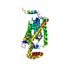
Mass: 42596.293 Da / Num. of mol.: 1 / Mutation: C-tail 1D4 epitope (ETSQVAPA)
Source method: isolated from a genetically manipulated source
Details: Residues 1-19, 257-262 and 336-380 are not modelled due to lack of electron density
Source: (gene. exp.) Hasarius adansoni (spider) / Gene: HaRh1 / Plasmid: pcDNA 3.0(+) / Details (production host): stable cell line / Cell line (production host): HEK293 GnTI- / Production host: Homo sapiens (human) / References: UniProt: B1B1U5
#2: Chemical ChemComp-RET / RETINAL


Mass: 284.436 Da / Num. of mol.: 1 / Source method: obtained synthetically / Formula: C20H28O / Feature type: SUBJECT OF INVESTIGATION
#3: Chemical
ChemComp-OLC / (2R)-2,3-dihydroxypropyl (9Z)-octadec-9-enoate / 1-Oleoyl-R-glycerol


Mass: 356.540 Da / Num. of mol.: 11 / Source method: obtained synthetically / Formula: C21H40O4
#4: Water ChemComp-HOH / water


Mass: 18.015 Da / Num. of mol.: 54 / Source method: isolated from a natural source / Formula: H2O

-
Experimental details

-
Experiment

ExperimentMethod: X-RAY DIFFRACTION / Number of used crystals: 1

-
Sample preparation

CrystalDensity Matthews: 3.52 Å3/Da / Density % sol: 65.04 % / Description: Rectangular plate like crystals
Crystal growTemperature: 293 K / Method: lipidic cubic phase / pH: 6.5 / Details: PEG 400, Bis-Tris pH6.5 / Temp details: Crystallization set up at 4 then moved to 20

-
Data collection

DiffractionMean temperature: 100 K / Serial crystal experiment: N
Diffraction sourceSource: SYNCHROTRON / Site: SLS / Beamline: X06SA / Wavelength: 1 Å
DetectorType: DECTRIS EIGER X 16M / Detector: PIXEL / Date: Mar 3, 2018
RadiationProtocol: SINGLE WAVELENGTH / Monochromatic (M) / Laue (L): M / Scattering type: x-ray
Radiation wavelengthWavelength: 1 Å / Relative weight: 1
ReflectionResolution: 2.1→46.773 Å / Num. obs: 18992 / % possible obs: 90.6 % / Redundancy: 6.1 % / Biso Wilson estimate: 38.56 Å2 / CC1/2: 0.995 / Rmerge(I) obs: 0.142 / Rpim(I) all: 0.092 / Rrim(I) all: 0.17 / Net I/σ(I): 7.2
Reflection shellResolution: 2.1→2.39 Å / Redundancy: 3.5 % / Rmerge(I) obs: 0.748 / Num. unique obs: 938 / CC1/2: 0.537 / % possible all: 51.7

-
Processing

Software
NameVersionClassification
PHENIX1.13_2998refinement
PDB_EXTRACT3.24data extraction
XDSdata reduction
STARANISOdata scaling
PHENIX1.13_2998phasing
RefinementMethod to determine structure: MOLECULAR REPLACEMENT
Starting model: 2Z73
Resolution: 2.145→46.773 Å / SU ML: 0.3 / Cross valid method: FREE R-VALUE / σ(F): 1.34 / Phase error: 31.46
RfactorNum. reflection% reflectionSelection details
Rfree0.2554 1167 6.15 %Random Selection
Rwork0.2146 ---
obs0.2172 18977 69.74 %-
Solvent computationShrinkage radii: 0.9 Å / VDW probe radii: 1.11 Å
Displacement parametersBiso max: 90.69 Å2 / Biso mean: 39.1137 Å2 / Biso min: 20.86 Å2
Refinement stepCycle: final / Resolution: 2.145→46.773 Å
ProteinNucleic acidLigandSolventTotal
Num. atoms2398 0 153 54 2605
Biso mean--49.72 39.76 -
Num. residues----310
LS refinement shell

Refine-ID: X-RAY DIFFRACTION / Rfactor Rfree error: 0

Resolution (Å)Rfactor RfreeNum. reflection RfreeRfactor RworkNum. reflection Rwork% reflection obs (%)
2.145-2.24230.27849188
2.2423-2.36050.292945490
2.3605-2.50840.320885151647
2.5084-2.70210.3204201293093
2.7021-2.9740.26782030.21823189100
2.974-3.40420.25042080.2143317199
3.4042-4.28850.22022040.1923313698

+
About Yorodumi

-
News

-
Feb 9, 2022. New format data for meta-information of EMDB entries

New format data for meta-information of EMDB entries

  • Version 3 of the EMDB header file is now the official format.
  • The previous official version 1.9 will be removed from the archive.

Related info.:EMDB header

External links:wwPDB to switch to version 3 of the EMDB data model

-
Aug 12, 2020. Covid-19 info

Covid-19 info

URL: https://pdbjlvh1.pdbj.org/emnavi/covid19.php

New page: Covid-19 featured information page in EM Navigator.

Related info.:Covid-19 info / Mar 5, 2020. Novel coronavirus structure data

+
Mar 5, 2020. Novel coronavirus structure data

Novel coronavirus structure data

Related info.:Yorodumi Speices / Aug 12, 2020. Covid-19 info

External links:COVID-19 featured content - PDBj / Molecule of the Month (242):Coronavirus Proteases

+
Jan 31, 2019. EMDB accession codes are about to change! (news from PDBe EMDB page)

EMDB accession codes are about to change! (news from PDBe EMDB page)

  • The allocation of 4 digits for EMDB accession codes will soon come to an end. Whilst these codes will remain in use, new EMDB accession codes will include an additional digit and will expand incrementally as the available range of codes is exhausted. The current 4-digit format prefixed with “EMD-” (i.e. EMD-XXXX) will advance to a 5-digit format (i.e. EMD-XXXXX), and so on. It is currently estimated that the 4-digit codes will be depleted around Spring 2019, at which point the 5-digit format will come into force.
  • The EM Navigator/Yorodumi systems omit the EMD- prefix.

Related info.:Q: What is EMD? / ID/Accession-code notation in Yorodumi/EM Navigator

External links:EMDB Accession Codes are Changing Soon! / Contact to PDBj

+
Jul 12, 2017. Major update of PDB

Major update of PDB

  • wwPDB released updated PDB data conforming to the new PDBx/mmCIF dictionary.
  • This is a major update changing the version number from 4 to 5, and with Remediation, in which all the entries are updated.
  • In this update, many items about electron microscopy experimental information are reorganized (e.g. em_software).
  • Now, EM Navigator and Yorodumi are based on the updated data.

External links:wwPDB Remediation / Enriched Model Files Conforming to OneDep Data Standards Now Available in the PDB FTP Archive

-
Yorodumi

Thousand views of thousand structures

  • Yorodumi is a browser for structure data from EMDB, PDB, SASBDB, etc.
  • This page is also the successor to EM Navigator detail page, and also detail information page/front-end page for Omokage search.
  • The word "yorodu" (or yorozu) is an old Japanese word meaning "ten thousand". "mi" (miru) is to see.

Related info.:EMDB / PDB / SASBDB / Comparison of 3 databanks / Yorodumi Search / Aug 31, 2016. New EM Navigator & Yorodumi / Yorodumi Papers / Jmol/JSmol / Function and homology information / Changes in new EM Navigator and Yorodumi

Read more