- PDB-2ol2: High Resolution Structure of Native PCI in Space Group P21 -
+
Open data
ID or keywords:
Loading...
-
Basic information
Entry
Database: PDB / ID: 2ol2
Title
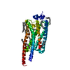
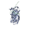
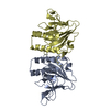
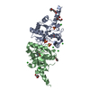
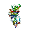
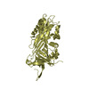
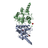
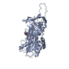
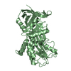
High Resolution Structure of Native PCI in Space Group P21
Components
Plasma serine protease inhibitor
Keywords
hydrolase inhibitor / serpin
Function / homology
Function and homology information
protein C inhibitor-TMPRSS7 complex / protein C inhibitor-TMPRSS11E complex / protein C inhibitor-PLAT complex / protein C inhibitor-PLAU complex / protein C inhibitor-thrombin complex / protein C inhibitor-KLK3 complex / protein C inhibitor-plasma kallikrein complex / seminal clot liquefaction / protein C inhibitor-coagulation factor V complex / protein C inhibitor-coagulation factor Xa complex ...protein C inhibitor-TMPRSS7 complex / protein C inhibitor-TMPRSS11E complex / protein C inhibitor-PLAT complex / protein C inhibitor-PLAU complex / protein C inhibitor-thrombin complex / protein C inhibitor-KLK3 complex / protein C inhibitor-plasma kallikrein complex / seminal clot liquefaction / protein C inhibitor-coagulation factor V complex / protein C inhibitor-coagulation factor Xa complex / protein C inhibitor-coagulation factor XI complex / acrosin binding / platelet dense tubular network / fusion of sperm to egg plasma membrane involved in single fertilization / acrosomal membrane / platelet alpha granule / glycosaminoglycan binding / phosphatidylcholine binding / retinoic acid binding / lipid transport / : / : / serine-type endopeptidase inhibitor activity / heparin binding / protease binding / spermatogenesis / external side of plasma membrane / protein-containing complex / : / extracellular exosome / extracellular region / membrane Similarity search - Function
In the structure databanks used in Yorodumi, some data are registered as the other names, "COVID-19 virus" and "2019-nCoV". Here are the details of the virus and the list of structure data.
Jan 31, 2019. EMDB accession codes are about to change! (news from PDBe EMDB page)
EMDB accession codes are about to change! (news from PDBe EMDB page)
The allocation of 4 digits for EMDB accession codes will soon come to an end. Whilst these codes will remain in use, new EMDB accession codes will include an additional digit and will expand incrementally as the available range of codes is exhausted. The current 4-digit format prefixed with “EMD-” (i.e. EMD-XXXX) will advance to a 5-digit format (i.e. EMD-XXXXX), and so on. It is currently estimated that the 4-digit codes will be depleted around Spring 2019, at which point the 5-digit format will come into force.
The EM Navigator/Yorodumi systems omit the EMD- prefix.
Related info.:Q: What is EMD? / ID/Accession-code notation in Yorodumi/EM Navigator
Yorodumi is a browser for structure data from EMDB, PDB, SASBDB, etc.
This page is also the successor to EM Navigator detail page, and also detail information page/front-end page for Omokage search.
The word "yorodu" (or yorozu) is an old Japanese word meaning "ten thousand". "mi" (miru) is to see.
Related info.:EMDB / PDB / SASBDB / Comparison of 3 databanks / Yorodumi Search / Aug 31, 2016. New EM Navigator & Yorodumi / Yorodumi Papers / Jmol/JSmol / Function and homology information / Changes in new EM Navigator and Yorodumi
 Movie
Movie Controller


 Open data
 Basic information
 Components
 Keywords
 Function and homology information
 Homo sapiens (human)
 X-RAY DIFFRACTION /
  Authors
 Citation
 Structure visualization
 Downloads & links
 Other downloads
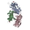


 PDBj
PDBj


 Assembly
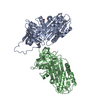


 Mass: 92.094 Da / Num. of mol.: 1 / Source method: obtained synthetically / Formula: C3H8O3
 Mass: 18.015 Da / Num. of mol.: 284 / Source method: isolated from a natural source / Formula: H2O
 Sample preparation
 / Beamline: ID14-3 / Wavelength: 0.931 Å
 Processing