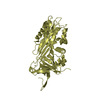
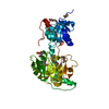
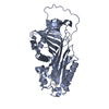
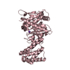
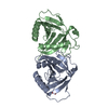
[English] 日本語
Yorodumi
- PDB-5nbv: Crystal structure of native alpha-1-antitrypsin with seven stabil... -

+
Open data


ID or keywords:

Loading...

-
Basic information

Entry
Database: PDB / ID: 5nbv
TitleCrystal structure of native alpha-1-antitrypsin with seven stabilising mutations
ComponentsAlpha-1-antitrypsin
KeywordsHYDROLASE / SERPIN
Function / homology
Function and homology information


Cargo concentration in the ER / COPII-mediated vesicle transport / COPII-coated ER to Golgi transport vesicle / endoplasmic reticulum-Golgi intermediate compartment membrane / platelet alpha granule lumen / acute-phase response / Post-translational protein phosphorylation / serine-type endopeptidase inhibitor activity / Regulation of Insulin-like Growth Factor (IGF) transport and uptake by Insulin-like Growth Factor Binding Proteins (IGFBPs) / blood coagulation ...Cargo concentration in the ER / COPII-mediated vesicle transport / COPII-coated ER to Golgi transport vesicle / endoplasmic reticulum-Golgi intermediate compartment membrane / platelet alpha granule lumen / acute-phase response / Post-translational protein phosphorylation / serine-type endopeptidase inhibitor activity / Regulation of Insulin-like Growth Factor (IGF) transport and uptake by Insulin-like Growth Factor Binding Proteins (IGFBPs) / blood coagulation / Platelet degranulation / protease binding / collagen-containing extracellular matrix / ficolin-1-rich granule lumen / endoplasmic reticulum lumen / intracellular membrane-bounded organelle / Neutrophil degranulation / Golgi apparatus / endoplasmic reticulum / extracellular space / extracellular exosome / extracellular region / identical protein binding
Similarity search - Function
Antithrombin; Chain I, domain 2 / Antithrombin, subunit I, domain 2 / Alpha-1-antitrypsin; domain 1 / Alpha-1-antitrypsin, domain 1 / Serpin, conserved site / Serpins signature. / Serpin superfamily, domain 2 / Serpin family / Serpin domain / Serpin superfamily ...Antithrombin; Chain I, domain 2 / Antithrombin, subunit I, domain 2 / Alpha-1-antitrypsin; domain 1 / Alpha-1-antitrypsin, domain 1 / Serpin, conserved site / Serpins signature. / Serpin superfamily, domain 2 / Serpin family / Serpin domain / Serpin superfamily / Serpin superfamily, domain 1 / Serpin (serine protease inhibitor) / SERine Proteinase INhibitors / Roll / 2-Layer Sandwich / Mainly Beta / Alpha Beta
Similarity search - Domain/homology
Biological speciesHomo sapiens (human)
MethodX-RAY DIFFRACTION / SYNCHROTRON / MOLECULAR REPLACEMENT / Resolution: 1.73 Å
AuthorsHuntington, J.A. / Pomowski, A. / Johnson, D.J.D.
Funding support United Kingdom, 1items
OrganizationGrant numberCountry
Medical Research Council (United Kingdom)MR/L017431/1 United Kingdom
CitationJournal: To Be Published
Title: CRYSTAL STRUCTURE OF THE Z VARIANT OF ALPHA-1-ANTITRYPSIN REVEALS STRUCTURAL AND DYNAMICAL CHANGES AND SUPPORTS A C-TERMINAL DOMAIN SWAP MECHANISM OF POLYMERIZATION
Authors: Johnson, D.J.D. / Pomowski, A. / Huntington, J.A.
History
DepositionMar 2, 2017Deposition site: PDBE / Processing site: PDBE
Revision 1.0Mar 21, 2018Provider: repository / Type: Initial release
Revision 1.1Oct 24, 2018Group: Data collection / Derived calculations / Category: struct_conn
Revision 1.2Oct 16, 2019Group: Data collection / Category: reflns_shell
Revision 1.3Jan 17, 2024Group: Advisory / Data collection ...Advisory / Data collection / Database references / Refinement description
Category: chem_comp_atom / chem_comp_bond ...chem_comp_atom / chem_comp_bond / database_2 / pdbx_initial_refinement_model / pdbx_unobs_or_zero_occ_atoms / pdbx_unobs_or_zero_occ_residues
Item: _database_2.pdbx_DOI / _database_2.pdbx_database_accession

-
Structure visualization

Structure viewerMolecule:
MolmilJmol/JSmol

Downloads & links

-
Assembly

Deposited unit
A: Alpha-1-antitrypsin
hetero molecules


Theoretical massNumber of molelcules
Total (without water)42,6174
Polymers42,3401
Non-polymers2763
Water3,603200
1


  • Idetical with deposited unit
  • defined by author&software
TypeNameSymmetry operationNumber
identity operation1_555x,y,z1
Buried area650 Å2
ΔGint-1 kcal/mol
Surface area15940 Å2
MethodPISA
Unit cell
Length a, b, c (Å)113.700, 39.260, 90.560
Angle α, β, γ (deg.)90.00, 104.08, 90.00
Int Tables number5
Space group name H-MC121

-
Components

#1: Protein Alpha-1-antitrypsin / Alpha-1 protease inhibitor / Alpha-1-antiproteinase / Serpin A1


Mass: 42340.375 Da / Num. of mol.: 1
Source method: isolated from a genetically manipulated source
Source: (gene. exp.) Homo sapiens (human) / Gene: SERPINA1, AAT, PI, PRO0684, PRO2209 / Production host: Escherichia coli (E. coli) / References: UniProt: P01009
#2: Chemical ChemComp-GOL / GLYCEROL / GLYCERIN / PROPANE-1,2,3-TRIOL


Mass: 92.094 Da / Num. of mol.: 3 / Source method: obtained synthetically / Formula: C3H8O3
#3: Water ChemComp-HOH / water


Mass: 18.015 Da / Num. of mol.: 200 / Source method: isolated from a natural source / Formula: H2O

-
Experimental details

-
Experiment

ExperimentMethod: X-RAY DIFFRACTION / Number of used crystals: 1

-
Sample preparation

CrystalDensity Matthews: 2.32 Å3/Da / Density % sol: 46.87 %
Crystal growTemperature: 293 K / Method: vapor diffusion, sitting drop / Details: PEG 4000, carboxylic acid mixture

-
Data collection

DiffractionMean temperature: 100 K
Diffraction sourceSource: SYNCHROTRON / Site: Diamond / Beamline: I02 / Wavelength: 0.9763 Å
DetectorType: DECTRIS PILATUS 6M-F / Detector: PIXEL / Date: Feb 25, 2013
RadiationProtocol: SINGLE WAVELENGTH / Monochromatic (M) / Laue (L): M / Scattering type: x-ray
Radiation wavelengthWavelength: 0.9763 Å / Relative weight: 1
ReflectionResolution: 1.73→52.85 Å / Num. obs: 40844 / % possible obs: 99.8 % / Redundancy: 3.1 % / Net I/σ(I): 7.4

-
Processing

Software
NameVersionClassification
REFMAC5.8.0155refinement
iMOSFLMdata reduction
Aimlessdata scaling
PHASERphasing
RefinementMethod to determine structure: MOLECULAR REPLACEMENT
Starting model: 5NBU
Resolution: 1.73→52.85 Å / Cor.coef. Fo:Fc: 0.958 / Cor.coef. Fo:Fc free: 0.938 / SU B: 4.654 / SU ML: 0.136 / Cross valid method: THROUGHOUT / ESU R: 0.132 / ESU R Free: 0.13 / Details: HYDROGENS HAVE BEEN ADDED IN THE RIDING POSITIONS
RfactorNum. reflection% reflectionSelection details
Rfree0.25931 2056 5 %RANDOM
Rwork0.21645 ---
obs0.21865 38771 99.61 %-
Solvent computationIon probe radii: 0.8 Å / Shrinkage radii: 0.8 Å / VDW probe radii: 1.2 Å
Displacement parametersBiso mean: 33.987 Å2
Baniso -1Baniso -2Baniso -3
1--0.9 Å20 Å21.4 Å2
2---0.4 Å20 Å2
3---0.52 Å2
Refinement stepCycle: 1 / Resolution: 1.73→52.85 Å
ProteinNucleic acidLigandSolventTotal
Num. atoms2789 0 18 200 3007
Refine LS restraints
Refine-IDTypeDev idealDev ideal targetNumber
X-RAY DIFFRACTIONr_bond_refined_d0.0020.0192989
X-RAY DIFFRACTIONr_bond_other_d00.022816
X-RAY DIFFRACTIONr_angle_refined_deg0.5811.974071
X-RAY DIFFRACTIONr_angle_other_deg0.45636463
X-RAY DIFFRACTIONr_dihedral_angle_1_deg11.4925391
X-RAY DIFFRACTIONr_dihedral_angle_2_deg37.10825.424118
X-RAY DIFFRACTIONr_dihedral_angle_3_deg13.46115473
X-RAY DIFFRACTIONr_dihedral_angle_4_deg21.095155
X-RAY DIFFRACTIONr_chiral_restr0.0440.2482
X-RAY DIFFRACTIONr_gen_planes_refined0.0030.0213421
X-RAY DIFFRACTIONr_gen_planes_other00.02658
X-RAY DIFFRACTIONr_nbd_refined
X-RAY DIFFRACTIONr_nbd_other
X-RAY DIFFRACTIONr_nbtor_refined
X-RAY DIFFRACTIONr_nbtor_other
X-RAY DIFFRACTIONr_xyhbond_nbd_refined
X-RAY DIFFRACTIONr_xyhbond_nbd_other
X-RAY DIFFRACTIONr_metal_ion_refined
X-RAY DIFFRACTIONr_metal_ion_other
X-RAY DIFFRACTIONr_symmetry_vdw_refined
X-RAY DIFFRACTIONr_symmetry_vdw_other
X-RAY DIFFRACTIONr_symmetry_hbond_refined
X-RAY DIFFRACTIONr_symmetry_hbond_other
X-RAY DIFFRACTIONr_symmetry_metal_ion_refined
X-RAY DIFFRACTIONr_symmetry_metal_ion_other
X-RAY DIFFRACTIONr_mcbond_it4.0463.3761552
X-RAY DIFFRACTIONr_mcbond_other4.0413.3731551
X-RAY DIFFRACTIONr_mcangle_it5.4285.031947
X-RAY DIFFRACTIONr_mcangle_other5.4275.0321948
X-RAY DIFFRACTIONr_scbond_it4.7173.681437
X-RAY DIFFRACTIONr_scbond_other4.7163.681437
X-RAY DIFFRACTIONr_scangle_it
X-RAY DIFFRACTIONr_scangle_other6.6615.382125
X-RAY DIFFRACTIONr_long_range_B_refined8.43340.5393196
X-RAY DIFFRACTIONr_long_range_B_other8.43340.5553197
X-RAY DIFFRACTIONr_rigid_bond_restr
X-RAY DIFFRACTIONr_sphericity_free
X-RAY DIFFRACTIONr_sphericity_bonded
LS refinement shellResolution: 1.73→1.775 Å / Total num. of bins used: 20
RfactorNum. reflection% reflection
Rfree0.365 160 -
Rwork0.37 2845 -
obs--99.5 %

+
About Yorodumi

-
News

-
Feb 9, 2022. New format data for meta-information of EMDB entries

New format data for meta-information of EMDB entries

  • Version 3 of the EMDB header file is now the official format.
  • The previous official version 1.9 will be removed from the archive.

Related info.:EMDB header

External links:wwPDB to switch to version 3 of the EMDB data model

-
Aug 12, 2020. Covid-19 info

Covid-19 info

URL: https://pdbjlc1.pdbj.org/emnavi/covid19.php

New page: Covid-19 featured information page in EM Navigator.

Related info.:Covid-19 info / Mar 5, 2020. Novel coronavirus structure data

+
Mar 5, 2020. Novel coronavirus structure data

Novel coronavirus structure data

Related info.:Yorodumi Speices / Aug 12, 2020. Covid-19 info

External links:COVID-19 featured content - PDBj / Molecule of the Month (242):Coronavirus Proteases

+
Jan 31, 2019. EMDB accession codes are about to change! (news from PDBe EMDB page)

EMDB accession codes are about to change! (news from PDBe EMDB page)

  • The allocation of 4 digits for EMDB accession codes will soon come to an end. Whilst these codes will remain in use, new EMDB accession codes will include an additional digit and will expand incrementally as the available range of codes is exhausted. The current 4-digit format prefixed with “EMD-” (i.e. EMD-XXXX) will advance to a 5-digit format (i.e. EMD-XXXXX), and so on. It is currently estimated that the 4-digit codes will be depleted around Spring 2019, at which point the 5-digit format will come into force.
  • The EM Navigator/Yorodumi systems omit the EMD- prefix.

Related info.:Q: What is EMD? / ID/Accession-code notation in Yorodumi/EM Navigator

External links:EMDB Accession Codes are Changing Soon! / Contact to PDBj

+
Jul 12, 2017. Major update of PDB

Major update of PDB

  • wwPDB released updated PDB data conforming to the new PDBx/mmCIF dictionary.
  • This is a major update changing the version number from 4 to 5, and with Remediation, in which all the entries are updated.
  • In this update, many items about electron microscopy experimental information are reorganized (e.g. em_software).
  • Now, EM Navigator and Yorodumi are based on the updated data.

External links:wwPDB Remediation / Enriched Model Files Conforming to OneDep Data Standards Now Available in the PDB FTP Archive

-
Yorodumi

Thousand views of thousand structures

  • Yorodumi is a browser for structure data from EMDB, PDB, SASBDB, etc.
  • This page is also the successor to EM Navigator detail page, and also detail information page/front-end page for Omokage search.
  • The word "yorodu" (or yorozu) is an old Japanese word meaning "ten thousand". "mi" (miru) is to see.

Related info.:EMDB / PDB / SASBDB / Comparison of 3 databanks / Yorodumi Search / Aug 31, 2016. New EM Navigator & Yorodumi / Yorodumi Papers / Jmol/JSmol / Function and homology information / Changes in new EM Navigator and Yorodumi

Read more