 Movie
Movie Controller

[English] 日本語
 Yorodumi
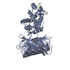
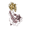
- PDB-6ys9: T_926 truncate of ChlH from Thermosynechococcus elongatus at 1.64... -
+ Open data

- Basic information
| Entry | Database: PDB / ID: 6ys9 | ||||||
|---|---|---|---|---|---|---|---|
| Title | T_926 truncate of ChlH from Thermosynechococcus elongatus at 1.64 A resolution | ||||||
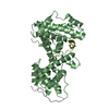
 Components Components | Magnesium-protoporphyrin methyltransferase | ||||||
 Keywords Keywords | PHOTOSYNTHESIS / Magnesium chelatase / chlorophyll / bacteriochlorophyll | ||||||
| Function / homology |  Function and homology information Function and homology informationmagnesium chelatase / magnesium chelatase activity / chlorophyll biosynthetic process / photosynthesis / methyltransferase activity / methylation / ATP binding Similarity search - Function | ||||||
| Biological species |   Thermosynechococcus elongatus (bacteria) Thermosynechococcus elongatus (bacteria) | ||||||
| Method |  X-RAY DIFFRACTION / SYNCHROTRON / MOLECULAR REPLACEMENT / Resolution: 1.64 Å X-RAY DIFFRACTION / SYNCHROTRON / MOLECULAR REPLACEMENT / Resolution: 1.64 Å | ||||||
 Authors Authors | Bisson, C. / Hunter, C.N. | ||||||
| Funding support | 1items
| ||||||
 Citation Citation | Journal: Nat.Plants / Year: 2020 Title: The active site of magnesium chelatase. Authors: Adams, N.B.P. / Bisson, C. / Brindley, A.A. / Farmer, D.A. / Davison, P.A. / Reid, J.D. / Hunter, C.N. | ||||||
| History |
|
- Structure visualization
| Structure viewer | Molecule: MolmilJmol/JSmol |
|---|
- Downloads & links
-Download
| PDBx/mmCIF format | 6ys9.cif.gz | 312.9 KB | Display | PDBx/mmCIF format |
|---|---|---|---|---|
| PDB format | pdb6ys9.ent.gz | 253.8 KB | Display | PDB format |
| PDBx/mmJSON format | 6ys9.json.gz | Tree view | PDBx/mmJSON format | |
| Others |  Other downloads Other downloads |
-Validation report
| Arichive directory | https://data.pdbj.org/pub/pdb/validation_reports/ys/6ys9ftp://data.pdbj.org/pub/pdb/validation_reports/ys/6ys9 | HTTPS FTP |
|---|
-Related structure data


-Links
 PDBj
PDBj- Assembly
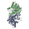
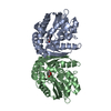
| Deposited unit | 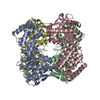
| ||||||||
|---|---|---|---|---|---|---|---|---|---|
| 1 | 
| ||||||||
| 2 | 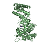
| ||||||||
| 3 | 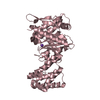
| ||||||||
| 4 | 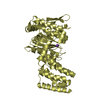
| ||||||||
| Unit cell |
|
-Components
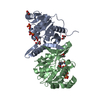
| #1: Protein | Mass: 46093.746 Da / Num. of mol.: 4 / Mutation: Truncated at the N-terminus Source method: isolated from a genetically manipulated source Details: C-terminal His-tag Source: (gene. exp.) Thermosynechococcus elongatus (strain BP-1) (bacteria)Gene: chlH / Production host: #2: Chemical | ChemComp-K /   Mass: 39.098 Da / Num. of mol.: 4 / Source method: obtained synthetically / Formula: K Mass: 39.098 Da / Num. of mol.: 4 / Source method: obtained synthetically / Formula: K#3: Water | ChemComp-HOH / |  Mass: 18.015 Da / Num. of mol.: 554 / Source method: isolated from a natural source / Formula: H2O Mass: 18.015 Da / Num. of mol.: 554 / Source method: isolated from a natural source / Formula: H2OHas ligand of interest | N | |
|---|
-Experimental details
-Experiment
| Experiment | Method: X-RAY DIFFRACTION / Number of used crystals: 1 |
|---|
- Sample preparation
| Crystal | Density Matthews: 2.15 Å3/Da / Density % sol: 42.82 % |
|---|---|
| Crystal grow | Temperature: 290 K / Method: vapor diffusion, sitting drop / pH: 8 / Details: 20-22% PEG 6000, 100 mM Tris pH 8, 200 mM MgCl2 |
-Data collection
| Diffraction | Mean temperature: 100 K / Serial crystal experiment: N | |||||||||||||||
|---|---|---|---|---|---|---|---|---|---|---|---|---|---|---|---|---|
| Diffraction source | Source: SYNCHROTRON / Site: Diamond  / Beamline: I03 / Wavelength: 0.97633 Å / Beamline: I03 / Wavelength: 0.97633 Å | |||||||||||||||
| Detector | Type: DECTRIS PILATUS3 S 6M / Detector: PIXEL / Date: Aug 8, 2015 | |||||||||||||||
| Radiation | Protocol: SINGLE WAVELENGTH / Monochromatic (M) / Laue (L): M / Scattering type: x-ray | |||||||||||||||
| Radiation wavelength | Wavelength: 0.97633 Å / Relative weight: 1 | |||||||||||||||
| Reflection twin |
| |||||||||||||||
| Reflection | Resolution: 1.64→64.38 Å / Num. obs: 172739 / % possible obs: 91.2 % / Redundancy: 2.3 % / CC1/2: 0.994 / Rmerge(I) obs: 0.073 / Rpim(I) all: 0.057 / Rrim(I) all: 0.093 / Net I/σ(I): 7.4 | |||||||||||||||
| Reflection shell | Resolution: 1.64→1.68 Å / Redundancy: 2.2 % / Rmerge(I) obs: 0.544 / Num. unique obs: 10643 / CC1/2: 0.487 / Rpim(I) all: 0.442 / Rrim(I) all: 0.704 / % possible all: 76.3 |
- Processing
| Software |
| ||||||||||||||||||||||||||||||||||||||||||||||||||||||||||||
|---|---|---|---|---|---|---|---|---|---|---|---|---|---|---|---|---|---|---|---|---|---|---|---|---|---|---|---|---|---|---|---|---|---|---|---|---|---|---|---|---|---|---|---|---|---|---|---|---|---|---|---|---|---|---|---|---|---|---|---|---|---|
| Refinement | Method to determine structure: MOLECULAR REPLACEMENT Starting model: SeMet poly-Ala model Resolution: 1.64→64.38 Å / Cor.coef. Fo:Fc: 0.943 / Cor.coef. Fo:Fc free: 0.918 / SU B: 1.91 / SU ML: 0.07 / Cross valid method: THROUGHOUT / σ(F): 0 / ESU R: 0.024 / ESU R Free: 0.025 Details: HYDROGENS HAVE BEEN ADDED IN THE RIDING POSITIONS U VALUES : REFINED INDIVIDUALLY
| ||||||||||||||||||||||||||||||||||||||||||||||||||||||||||||
| Solvent computation | Ion probe radii: 0.8 Å / Shrinkage radii: 0.8 Å / VDW probe radii: 1.2 Å | ||||||||||||||||||||||||||||||||||||||||||||||||||||||||||||
| Displacement parameters | Biso max: 200 Å2 / Biso mean: 24.569 Å2 / Biso min: 2.32 Å2
| ||||||||||||||||||||||||||||||||||||||||||||||||||||||||||||
| Refinement step | Cycle: final / Resolution: 1.64→64.38 Å
| ||||||||||||||||||||||||||||||||||||||||||||||||||||||||||||
| Refine LS restraints |
| ||||||||||||||||||||||||||||||||||||||||||||||||||||||||||||
| LS refinement shell | Resolution: 1.611→1.653 Å / Rfactor Rfree error: 0 / Total num. of bins used: 20
|
