 Movie
Movie Controller

[English] 日本語
 Yorodumi
- PDB-4i2y: Crystal Structure of the genetically encoded calcium indicator RGECO1 -
+ Open data

- Basic information
| Entry | Database: PDB / ID: 4i2y | ||||||
|---|---|---|---|---|---|---|---|
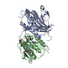
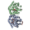
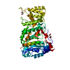
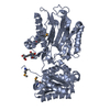
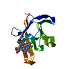
| Title | Crystal Structure of the genetically encoded calcium indicator RGECO1 | ||||||
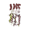
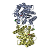
 Components Components | RGECO1 | ||||||
 Keywords Keywords | FLUORESCENT PROTEIN / Calcium binding / sensor / mApple / fluorescent calcium indicator | ||||||
| Function / homology |  Function and homology information Function and homology informationregulation of store-operated calcium channel activity / : / DAPK1-calmodulin complex / : / regulation of response to tumor cell / positive regulation of autophagic cell death / : / : / : / : ...regulation of store-operated calcium channel activity / : / DAPK1-calmodulin complex / : / regulation of response to tumor cell / positive regulation of autophagic cell death / : / : / : / : / : / : / type 3 metabotropic glutamate receptor binding / establishment of protein localization to membrane / positive regulation of DNA binding / negative regulation of high voltage-gated calcium channel activity / negative regulation of ryanodine-sensitive calcium-release channel activity / organelle localization by membrane tethering / : / autophagosome membrane docking / regulation of synaptic vesicle exocytosis / negative regulation of calcium ion export across plasma membrane / regulation of cardiac muscle cell action potential / nitric-oxide synthase binding / adenylate cyclase binding / protein phosphatase activator activity / regulation of ryanodine-sensitive calcium-release channel activity / regulation of synaptic vesicle endocytosis / detection of calcium ion / catalytic complex / regulation of cardiac muscle contraction / positive regulation of nitric-oxide synthase activity / phosphatidylinositol 3-kinase binding / activation of adenylate cyclase activity / calcium channel inhibitor activity / regulation of release of sequestered calcium ion into cytosol by sarcoplasmic reticulum / enzyme regulator activity / regulation of calcium-mediated signaling / titin binding / voltage-gated potassium channel complex / calcium channel complex / potassium ion transmembrane transport / regulation of heart rate / response to amphetamine / nitric-oxide synthase regulator activity / adenylate cyclase activator activity / bioluminescence / regulation of cytokinesis / spindle microtubule / sarcomere / calcium-mediated signaling / generation of precursor metabolites and energy / calcium channel regulator activity / response to calcium ion / G2/M transition of mitotic cell cycle / spindle pole / disordered domain specific binding / calcium-dependent protein binding / kinase activity / protein autophosphorylation / myelin sheath / growth cone / vesicle / transmembrane transporter binding / calmodulin binding / neuron projection / positive regulation of apoptotic process / protein domain specific binding / calcium ion binding / centrosome / protein kinase binding / protein-containing complex / mitochondrion / nucleoplasm / nucleus / plasma membrane / cytosol / cytoplasm Similarity search - Function | ||||||
| Biological species |   Discosoma sp. (sea anemone) Discosoma sp. (sea anemone) | ||||||
| Method |  X-RAY DIFFRACTION / SYNCHROTRON / MOLECULAR REPLACEMENT / Resolution: 2.2 Å X-RAY DIFFRACTION / SYNCHROTRON / MOLECULAR REPLACEMENT / Resolution: 2.2 Å | ||||||
 Authors Authors | Akerboom, J. / Looger, L.L. / Schreiter, E.R. | ||||||
 Citation Citation | Journal: Front Mol Neurosci / Year: 2013 Title: Genetically encoded calcium indicators for multi-color neural activity imaging and combination with optogenetics. Authors: Akerboom, J. / Carreras Calderon, N. / Tian, L. / Wabnig, S. / Prigge, M. / Tolo, J. / Gordus, A. / Orger, M.B. / Severi, K.E. / Macklin, J.J. / Patel, R. / Pulver, S.R. / Wardill, T.J. / ...Authors: Akerboom, J. / Carreras Calderon, N. / Tian, L. / Wabnig, S. / Prigge, M. / Tolo, J. / Gordus, A. / Orger, M.B. / Severi, K.E. / Macklin, J.J. / Patel, R. / Pulver, S.R. / Wardill, T.J. / Fischer, E. / Schuler, C. / Chen, T.W. / Sarkisyan, K.S. / Marvin, J.S. / Bargmann, C.I. / Kim, D.S. / Kugler, S. / Lagnado, L. / Hegemann, P. / Gottschalk, A. / Schreiter, E.R. / Looger, L.L. | ||||||
| History |
|
- Structure visualization
| Structure viewer | Molecule: MolmilJmol/JSmol |
|---|
- Downloads & links
-Download
| PDBx/mmCIF format | 4i2y.cif.gz | 171.2 KB | Display | PDBx/mmCIF format |
|---|---|---|---|---|
| PDB format | pdb4i2y.ent.gz | 131.6 KB | Display | PDB format |
| PDBx/mmJSON format | 4i2y.json.gz | Tree view | PDBx/mmJSON format | |
| Others |  Other downloads Other downloads |
-Validation report
| Arichive directory | https://data.pdbj.org/pub/pdb/validation_reports/i2/4i2yftp://data.pdbj.org/pub/pdb/validation_reports/i2/4i2y | HTTPS FTP |
|---|
-Related structure data
| Related structure data |  3u0kC  3u0lC 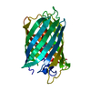 3u0mC  3u0nC  2h5qS 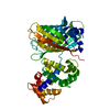 3sg3S S: Starting model for refinement C: citing same article ( |
|---|---|
| Similar structure data |


-Links
 PDBj
PDBj


- Assembly
| Deposited unit | 
| ||||||||
|---|---|---|---|---|---|---|---|---|---|
| 1 | 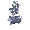
| ||||||||
| 2 | 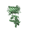
| ||||||||
| Unit cell |
|
-Components
| #1: Protein | Mass: 47530.219 Da / Num. of mol.: 2 Mutation: Q44N, T46A, T62S, L65M, R68E, V71A, G74S, H77K, A79G, K81R, L89A, V90A, F92V, S94T, I95T, M97K, Y107A, Y109I, S112I, T117V, Y129C, T132A, R174H, T178S, G197A, H198F, N199Q, V201A, V228A, ...Mutation: Q44N, T46A, T62S, L65M, R68E, V71A, G74S, H77K, A79G, K81R, L89A, V90A, F92V, S94T, I95T, M97K, Y107A, Y109I, S112I, T117V, Y129C, T132A, R174H, T178S, G197A, H198F, N199Q, V201A, V228A, V230I, K240F, K249R, V261I, V262I, T263H, T265N, C274V, F281L, I282R, V284T, S288P, N363D, I366F, K380N, S404G, N414D, E430V Source method: isolated from a genetically manipulated source Source: (gene. exp.) Discosoma sp. (sea anemone), (gene. exp.) Production host:  References: UniProt: Q6LDG3, UniProt: Q9U6Y8, UniProt: P62161 #2: Chemical | ChemComp-CA /   Mass: 40.078 Da / Num. of mol.: 8 / Source method: obtained synthetically / Formula: Ca Mass: 40.078 Da / Num. of mol.: 8 / Source method: obtained synthetically / Formula: Ca#3: Water | ChemComp-HOH / |  Mass: 18.015 Da / Num. of mol.: 99 / Source method: isolated from a natural source / Formula: H2O Mass: 18.015 Da / Num. of mol.: 99 / Source method: isolated from a natural source / Formula: H2OSequence details | THIS TRIPLE CHIMERA COMPRISES (FROM THE N-TERMINUS) AN ENGINEERED EXPRESSION TAG, RESIDUES 37-55 OF ...THIS TRIPLE CHIMERA COMPRISES (FROM THE N-TERMINUS) AN ENGINEERED | |
|---|
-Experimental details
-Experiment
| Experiment | Method: X-RAY DIFFRACTION / Number of used crystals: 1 |
|---|
- Sample preparation
| Crystal | Density Matthews: 2.15 Å3/Da / Density % sol: 42.7 % |
|---|---|
| Crystal grow | Temperature: 277 K / Method: vapor diffusion, hanging drop / pH: 5 Details: 73mM malonic acid, 10mM ammonium citrate tribasic, 5mM succinic acid, 12mM malic acid, 16mM sodium acetate trihydrate, 20mM sodium formate, 6mM ammonium tartrate dibasic, 12% PEG 3350, pH 5, ...Details: 73mM malonic acid, 10mM ammonium citrate tribasic, 5mM succinic acid, 12mM malic acid, 16mM sodium acetate trihydrate, 20mM sodium formate, 6mM ammonium tartrate dibasic, 12% PEG 3350, pH 5, VAPOR DIFFUSION, HANGING DROP, temperature 277K |
-Data collection
| Diffraction | Mean temperature: 100 K |
|---|---|
| Diffraction source | Source: SYNCHROTRON / Site: ALS  / Beamline: 8.2.2 / Wavelength: 1 Å / Beamline: 8.2.2 / Wavelength: 1 Å |
| Detector | Type: ADSC QUANTUM 315 / Detector: CCD / Date: Nov 16, 2012 |
| Radiation | Monochromator: Double crystal, Si(111) / Protocol: SINGLE WAVELENGTH / Monochromatic (M) / Laue (L): M / Scattering type: x-ray |
| Radiation wavelength | Wavelength: 1 Å / Relative weight: 1 |
| Reflection | Resolution: 2.2→54.77 Å / Num. all: 40865 / Num. obs: 40865 / % possible obs: 100 % / Observed criterion σ(F): 0 / Observed criterion σ(I): 0 |
| Reflection shell | Resolution: 2.2→2.32 Å / % possible all: 100 |
- Processing
| Software |
| ||||||||||||||||||||||||||||||||||||||||||||||||||||||||||||||||||||||||||||||||||||||||||||||||||||||||||||||||||||||||||||||||||||||||||||||||||||||||||||||||||||||||||
|---|---|---|---|---|---|---|---|---|---|---|---|---|---|---|---|---|---|---|---|---|---|---|---|---|---|---|---|---|---|---|---|---|---|---|---|---|---|---|---|---|---|---|---|---|---|---|---|---|---|---|---|---|---|---|---|---|---|---|---|---|---|---|---|---|---|---|---|---|---|---|---|---|---|---|---|---|---|---|---|---|---|---|---|---|---|---|---|---|---|---|---|---|---|---|---|---|---|---|---|---|---|---|---|---|---|---|---|---|---|---|---|---|---|---|---|---|---|---|---|---|---|---|---|---|---|---|---|---|---|---|---|---|---|---|---|---|---|---|---|---|---|---|---|---|---|---|---|---|---|---|---|---|---|---|---|---|---|---|---|---|---|---|---|---|---|---|---|---|---|---|---|
| Refinement | Method to determine structure: MOLECULAR REPLACEMENT Starting model: 2H5Q, 3SG3 Resolution: 2.2→68.28 Å / Cor.coef. Fo:Fc: 0.947 / Cor.coef. Fo:Fc free: 0.909 / SU B: 6.21 / SU ML: 0.157 / Cross valid method: THROUGHOUT / ESU R: 0.288 / ESU R Free: 0.23 / Stereochemistry target values: MAXIMUM LIKELIHOOD / Details: HYDROGENS HAVE BEEN USED IF PRESENT IN THE INPUT
| ||||||||||||||||||||||||||||||||||||||||||||||||||||||||||||||||||||||||||||||||||||||||||||||||||||||||||||||||||||||||||||||||||||||||||||||||||||||||||||||||||||||||||
| Solvent computation | Ion probe radii: 0.8 Å / Shrinkage radii: 0.8 Å / VDW probe radii: 1.2 Å / Solvent model: MASK | ||||||||||||||||||||||||||||||||||||||||||||||||||||||||||||||||||||||||||||||||||||||||||||||||||||||||||||||||||||||||||||||||||||||||||||||||||||||||||||||||||||||||||
| Displacement parameters | Biso mean: 27.172 Å2
| ||||||||||||||||||||||||||||||||||||||||||||||||||||||||||||||||||||||||||||||||||||||||||||||||||||||||||||||||||||||||||||||||||||||||||||||||||||||||||||||||||||||||||
| Refinement step | Cycle: LAST / Resolution: 2.2→68.28 Å
| ||||||||||||||||||||||||||||||||||||||||||||||||||||||||||||||||||||||||||||||||||||||||||||||||||||||||||||||||||||||||||||||||||||||||||||||||||||||||||||||||||||||||||
| Refine LS restraints |
| ||||||||||||||||||||||||||||||||||||||||||||||||||||||||||||||||||||||||||||||||||||||||||||||||||||||||||||||||||||||||||||||||||||||||||||||||||||||||||||||||||||||||||
| LS refinement shell | Resolution: 2.2→2.257 Å / Total num. of bins used: 20
|
