 Movie
Movie Controller

[English] 日本語
 Yorodumi
- PDB-3apu: Crystal structure of the A variant of human alpha1-acid glycoprotein -
+ Open data

- Basic information
| Entry | Database: PDB / ID: 3apu | ||||||
|---|---|---|---|---|---|---|---|
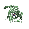
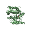
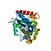
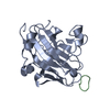
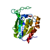
| Title | Crystal structure of the A variant of human alpha1-acid glycoprotein | ||||||
 Components Components | Alpha-1-acid glycoprotein 2 | ||||||
 Keywords Keywords | TRANSPORT PROTEIN / beta barrel / PLASMA PROTEIN | ||||||
| Function / homology |  Function and homology information Function and homology informationpositive regulation of interleukin-1 production / regulation of immune system process / platelet alpha granule lumen / acute-phase response / positive regulation of interleukin-1 beta production / specific granule lumen / positive regulation of tumor necrosis factor production / azurophil granule lumen / Platelet degranulation / blood microparticle ...positive regulation of interleukin-1 production / regulation of immune system process / platelet alpha granule lumen / acute-phase response / positive regulation of interleukin-1 beta production / specific granule lumen / positive regulation of tumor necrosis factor production / azurophil granule lumen / Platelet degranulation / blood microparticle / Neutrophil degranulation / : / extracellular exosome / extracellular region Similarity search - Function | ||||||
| Biological species |  Homo sapiens (human) Homo sapiens (human) | ||||||
| Method |  X-RAY DIFFRACTION / SYNCHROTRON / MOLECULAR REPLACEMENT / Resolution: 2.1 Å X-RAY DIFFRACTION / SYNCHROTRON / MOLECULAR REPLACEMENT / Resolution: 2.1 Å | ||||||
 Authors Authors | Nishi, K. / Ono, T. / Nakamura, T. / Fukunaga, N. / Izumi, M. / Watanabe, H. / Suenaga, A. / Maruyama, T. / Yamagata, Y. / Curry, S. / Otagiri, M. | ||||||
 Citation Citation | Journal: J. Biol. Chem. / Year: 2011 Title: Structural insights into differences in drug-binding selectivity between two forms of human alpha1-acid glycoprotein genetic variants, the A and F1*S forms. Authors: Nishi, K. / Ono, T. / Nakamura, T. / Fukunaga, N. / Izumi, M. / Watanabe, H. / Suenaga, A. / Maruyama, T. / Yamagata, Y. / Curry, S. / Otagiri, M. | ||||||
| History |
|
- Structure visualization
| Structure viewer | Molecule: MolmilJmol/JSmol |
|---|
- Downloads & links
-Download
| PDBx/mmCIF format | 3apu.cif.gz | 161.8 KB | Display | PDBx/mmCIF format |
|---|---|---|---|---|
| PDB format | pdb3apu.ent.gz | 128.6 KB | Display | PDB format |
| PDBx/mmJSON format | 3apu.json.gz | Tree view | PDBx/mmJSON format | |
| Others |  Other downloads Other downloads |
-Validation report
| Arichive directory | https://data.pdbj.org/pub/pdb/validation_reports/ap/3apuftp://data.pdbj.org/pub/pdb/validation_reports/ap/3apu | HTTPS FTP |
|---|
-Related structure data
| Related structure data |  3apvC  3apwC  3apxC  3bx6 C: citing same article ( S: Starting model for refinement |
|---|---|
| Similar structure data |


-Links
 PDBj
PDBj

- Assembly
| Deposited unit | 
| ||||||||
|---|---|---|---|---|---|---|---|---|---|
| 1 | 
| ||||||||
| 2 | 
| ||||||||
| Unit cell |
|
-Components
| #1: Protein | Mass: 22691.287 Da / Num. of mol.: 2 / Mutation: C149R Source method: isolated from a genetically manipulated source Source: (gene. exp.) Homo sapiens (human) / Gene: AGP2, ORM2 / Plasmid: pET3c / Production host:  #2: Chemical |   Mass: 194.226 Da / Num. of mol.: 2 / Source method: obtained synthetically / Formula: C8H18O5 / Comment: precipitant*YM Mass: 194.226 Da / Num. of mol.: 2 / Source method: obtained synthetically / Formula: C8H18O5 / Comment: precipitant*YM#3: Water | ChemComp-HOH / |  Mass: 18.015 Da / Num. of mol.: 83 / Source method: isolated from a natural source / Formula: H2O Mass: 18.015 Da / Num. of mol.: 83 / Source method: isolated from a natural source / Formula: H2OHas protein modification | Y | |
|---|
-Experimental details
-Experiment
| Experiment | Method: X-RAY DIFFRACTION / Number of used crystals: 1 |
|---|
- Sample preparation
| Crystal | Density Matthews: 2 Å3/Da / Density % sol: 38.56 % |
|---|---|
| Crystal grow | Temperature: 293 K / Method: vapor diffusion, hanging drop / pH: 4.6 Details: 30% PEG 4000, 0.2M ammonium acetate, 0.1M sodium acetate, pH 4.6, VAPOR DIFFUSION, HANGING DROP, temperature 293K |
-Data collection
| Diffraction | Mean temperature: 100 K |
|---|---|
| Diffraction source | Source: SYNCHROTRON / Site: SPring-8  / Beamline: BL44XU / Wavelength: 0.9 Å / Beamline: BL44XU / Wavelength: 0.9 Å |
| Detector | Type: Bruker DIP-6040 / Detector: CCD / Date: Jun 20, 2008 |
| Radiation | Protocol: SINGLE WAVELENGTH / Monochromatic (M) / Laue (L): M / Scattering type: x-ray |
| Radiation wavelength | Wavelength: 0.9 Å / Relative weight: 1 |
| Reflection | Resolution: 2.1→50 Å / Num. all: 21258 / Num. obs: 21258 / % possible obs: 99.9 % / Rmerge(I) obs: 0.044 / Net I/σ(I): 56.8 |
| Reflection shell | Resolution: 2.1→2.18 Å / Rmerge(I) obs: 0.326 / Mean I/σ(I) obs: 6.7 / % possible all: 99.3 |
- Processing
| Software |
| ||||||||||||||||||||||||||||||||||||||||||||||||||||||||||||||||||||||||||||||||||||||||||||||||||||||||||||||||||||||||||||||||||||||||||||||||||||||||||||||||||||||||||
|---|---|---|---|---|---|---|---|---|---|---|---|---|---|---|---|---|---|---|---|---|---|---|---|---|---|---|---|---|---|---|---|---|---|---|---|---|---|---|---|---|---|---|---|---|---|---|---|---|---|---|---|---|---|---|---|---|---|---|---|---|---|---|---|---|---|---|---|---|---|---|---|---|---|---|---|---|---|---|---|---|---|---|---|---|---|---|---|---|---|---|---|---|---|---|---|---|---|---|---|---|---|---|---|---|---|---|---|---|---|---|---|---|---|---|---|---|---|---|---|---|---|---|---|---|---|---|---|---|---|---|---|---|---|---|---|---|---|---|---|---|---|---|---|---|---|---|---|---|---|---|---|---|---|---|---|---|---|---|---|---|---|---|---|---|---|---|---|---|---|---|---|
| Refinement | Method to determine structure: MOLECULAR REPLACEMENT Starting model: PDB ENTRY 3BX6 3bx6 Resolution: 2.1→31.99 Å / Cor.coef. Fo:Fc: 0.959 / Cor.coef. Fo:Fc free: 0.947 / SU B: 14.626 / SU ML: 0.173 / Cross valid method: THROUGHOUT / ESU R: 0.257 / ESU R Free: 0.191 / Stereochemistry target values: MAXIMUM LIKELIHOOD / Details: HYDROGENS HAVE BEEN ADDED IN THE RIDING POSITIONS
| ||||||||||||||||||||||||||||||||||||||||||||||||||||||||||||||||||||||||||||||||||||||||||||||||||||||||||||||||||||||||||||||||||||||||||||||||||||||||||||||||||||||||||
| Solvent computation | Ion probe radii: 0.8 Å / Shrinkage radii: 0.8 Å / VDW probe radii: 1.4 Å / Solvent model: MASK | ||||||||||||||||||||||||||||||||||||||||||||||||||||||||||||||||||||||||||||||||||||||||||||||||||||||||||||||||||||||||||||||||||||||||||||||||||||||||||||||||||||||||||
| Displacement parameters | Biso mean: 51.507 Å2
| ||||||||||||||||||||||||||||||||||||||||||||||||||||||||||||||||||||||||||||||||||||||||||||||||||||||||||||||||||||||||||||||||||||||||||||||||||||||||||||||||||||||||||
| Refinement step | Cycle: LAST / Resolution: 2.1→31.99 Å
| ||||||||||||||||||||||||||||||||||||||||||||||||||||||||||||||||||||||||||||||||||||||||||||||||||||||||||||||||||||||||||||||||||||||||||||||||||||||||||||||||||||||||||
| Refine LS restraints |
| ||||||||||||||||||||||||||||||||||||||||||||||||||||||||||||||||||||||||||||||||||||||||||||||||||||||||||||||||||||||||||||||||||||||||||||||||||||||||||||||||||||||||||
| LS refinement shell | Resolution: 2.1→2.155 Å / Total num. of bins used: 20
| ||||||||||||||||||||||||||||||||||||||||||||||||||||||||||||||||||||||||||||||||||||||||||||||||||||||||||||||||||||||||||||||||||||||||||||||||||||||||||||||||||||||||||
| Refinement TLS params. | Method: refined / Refine-ID: X-RAY DIFFRACTION
| ||||||||||||||||||||||||||||||||||||||||||||||||||||||||||||||||||||||||||||||||||||||||||||||||||||||||||||||||||||||||||||||||||||||||||||||||||||||||||||||||||||||||||
| Refinement TLS group |
|
