 Movie
Movie Controller

[English] 日本語
 Yorodumi
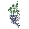
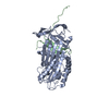
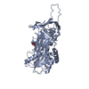
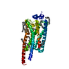
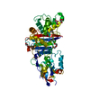
- PDB-4oak: Crystal structure of vancomycin resistance D,D-dipeptidase/D,D-pe... -
+ Open data

- Basic information
| Entry | Database: PDB / ID: 4oak | ||||||
|---|---|---|---|---|---|---|---|
| Title | Crystal structure of vancomycin resistance D,D-dipeptidase/D,D-pentapeptidase VanXYc D59S mutant in complex with D-Alanine-D-Alanine and copper (II) | ||||||
 Components Components | D,D-dipeptidase/D,D-carboxypeptidase | ||||||
 Keywords Keywords | HYDROLASE / CENTER FOR STRUCTURAL GENOMICS OF INFECTIOUS DISEASES / CSGID / NATIONAL INSTITUTE OF ALLERGY AND INFECTIOUS DISEASES / NIAID / ALPHA+BETA PROTEIN / METALLOPEPTIDASE / HEDGEHOG/DD-PEPTIDASE FOLD / MEROPS M15B SUBFAMILY / ZN2+-DEPENDENT / VANCOMYCIN RESISTANCE / ANTIBIOTIC RESISTANCE | ||||||
| Function / homology |  Function and homology information Function and homology informationD-Ala-D-Ala dipeptidase / D-Ala-D-Ala dipeptidase activity / Hydrolases; Acting on peptide bonds (peptidases); Metallocarboxypeptidases / carboxypeptidase activity / response to antibiotic / proteolysis / metal ion binding / cytoplasm Similarity search - Function | ||||||
| Biological species |  Enterococcus gallinarum (bacteria) Enterococcus gallinarum (bacteria) | ||||||
| Method |  X-RAY DIFFRACTION / MOLECULAR REPLACEMENT / Resolution: 2 Å X-RAY DIFFRACTION / MOLECULAR REPLACEMENT / Resolution: 2 Å | ||||||
 Authors Authors | Stogios, P.J. / Evdokimova, E. / Meziane-Cherif, D. / Di Leo, R. / Yim, V. / Courvalin, P. / Savchenko, A. / Anderson, W.F. / Center for Structural Genomics of Infectious Diseases (CSGID) | ||||||
 Citation Citation | Journal: Proc.Natl.Acad.Sci.USA / Year: 2014 Title: Structural basis for the evolution of vancomycin resistance D,D-peptidases. Authors: Meziane-Cherif, D. / Stogios, P.J. / Evdokimova, E. / Savchenko, A. / Courvalin, P. | ||||||
| History |
|
- Structure visualization
| Structure viewer | Molecule: MolmilJmol/JSmol |
|---|
- Downloads & links
-Download
| PDBx/mmCIF format | 4oak.cif.gz | 187 KB | Display | PDBx/mmCIF format |
|---|---|---|---|---|
| PDB format | pdb4oak.ent.gz | 146.6 KB | Display | PDB format |
| PDBx/mmJSON format | 4oak.json.gz | Tree view | PDBx/mmJSON format | |
| Others |  Other downloads Other downloads |
-Validation report
| Arichive directory | https://data.pdbj.org/pub/pdb/validation_reports/oa/4oakftp://data.pdbj.org/pub/pdb/validation_reports/oa/4oak | HTTPS FTP |
|---|
-Related structure data
| Related structure data | 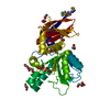 4f78C 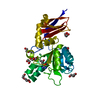 4muqC  4murSC 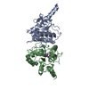 4musC  4mutC C: citing same article ( S: Starting model for refinement |
|---|---|
| Similar structure data | |
| Other databases |


-Links
 PDBj
PDBj
- Assembly
| Deposited unit | 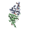
| ||||||||
|---|---|---|---|---|---|---|---|---|---|
| 1 |
| ||||||||
| Unit cell |
|
-Components
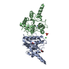
-Protein , 1 types, 2 molecules AB
| #1: Protein | Mass: 24765.953 Da / Num. of mol.: 2 / Fragment: VanXYc / Mutation: D59S Source method: isolated from a genetically manipulated source Source: (gene. exp.) Enterococcus gallinarum (bacteria) / Strain: BM4174 / Gene: vanXYc / Plasmid: P15TV-LIC / Production host: |
|---|
-Non-polymers , 7 types, 344 molecules 
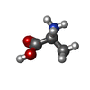


| #2: Chemical |  Mass: 63.546 Da / Num. of mol.: 2 / Source method: obtained synthetically / Formula: Cu Mass: 63.546 Da / Num. of mol.: 2 / Source method: obtained synthetically / Formula: Cu#3: Chemical | ChemComp-DAL /  Type: D-peptide linking / Mass: 89.093 Da / Num. of mol.: 4 / Source method: obtained synthetically / Formula: C3H7NO2 Type: D-peptide linking / Mass: 89.093 Da / Num. of mol.: 4 / Source method: obtained synthetically / Formula: C3H7NO2#4: Chemical |  Mass: 96.063 Da / Num. of mol.: 3 / Source method: obtained synthetically / Formula: SO4 Mass: 96.063 Da / Num. of mol.: 3 / Source method: obtained synthetically / Formula: SO4#5: Chemical | ChemComp-MG / |  Mass: 24.305 Da / Num. of mol.: 1 / Source method: obtained synthetically / Formula: Mg Mass: 24.305 Da / Num. of mol.: 1 / Source method: obtained synthetically / Formula: Mg#6: Chemical | ChemComp-CL /  Mass: 35.453 Da / Num. of mol.: 6 / Source method: obtained synthetically / Formula: Cl Mass: 35.453 Da / Num. of mol.: 6 / Source method: obtained synthetically / Formula: Cl#7: Chemical | ChemComp-PE3 /  Mass: 634.751 Da / Num. of mol.: 4 / Source method: obtained synthetically / Formula: C28H58O15 Mass: 634.751 Da / Num. of mol.: 4 / Source method: obtained synthetically / Formula: C28H58O15#8: Water | ChemComp-HOH / | Mass: 18.015 Da / Num. of mol.: 324 / Source method: isolated from a natural source / Formula: H2O |
|---|
-Details
| Has protein modification | Y |
|---|
-Experimental details
-Experiment
| Experiment | Method: X-RAY DIFFRACTION / Number of used crystals: 1 |
|---|
- Sample preparation
| Crystal | Density Matthews: 2.09 Å3/Da / Density % sol: 41.22 % |
|---|---|
| Crystal grow | Method: vapor diffusion, hanging drop / pH: 5.6 Details: 0.1 M MgCl2, 0.05 M MES, 20% PEG8K and 0.1 M phenantroline, 2 h incubation, transferred crystal to new drop with 0.1 M MgCl2, 0.05 M MES, 22% PEG8K and 1 mM CuCl2, 1.5 h incubation, ...Details: 0.1 M MgCl2, 0.05 M MES, 20% PEG8K and 0.1 M phenantroline, 2 h incubation, transferred crystal to new drop with 0.1 M MgCl2, 0.05 M MES, 22% PEG8K and 1 mM CuCl2, 1.5 h incubation, transferred crystal to new drop with 0.1 M MgCl2, 0.05 M MES, 22% PEG8K, 1 mM CuCl2 and 20 mM D-Ala-D-Ala. Cryoprotectant: paratone, pH 5.6, VAPOR DIFFUSION, HANGING DROP |
-Data collection
| Diffraction | Mean temperature: 100 K |
|---|---|
| Diffraction source | Source: ROTATING ANODE / Type: RIGAKU MICROMAX-007 HF / Wavelength: 1.5418 Å |
| Detector | Type: RIGAKU RAXIS IV++ / Detector: IMAGE PLATE / Date: Dec 12, 2013 |
| Radiation | Monochromator: graphite / Protocol: SINGLE WAVELENGTH / Monochromatic (M) / Laue (L): M / Scattering type: x-ray |
| Radiation wavelength | Wavelength: 1.5418 Å / Relative weight: 1 |
| Reflection | Resolution: 2→19.33 Å / Num. obs: 25760 / % possible obs: 94.9 % / Observed criterion σ(F): 0 / Observed criterion σ(I): -2 / Redundancy: 3.3 % / Rmerge(I) obs: 0.079 / Net I/σ(I): 11 |
| Reflection shell | Resolution: 2→2.11 Å / Redundancy: 3.3 % / Rmerge(I) obs: 0.518 / Mean I/σ(I) obs: 2.3 / % possible all: 92.3 |
- Processing
| Software |
| |||||||||||||||||||||||||||||||||||||||||||||||||||||||||||||||||||||||||||||||||||||||||||||||||||||||||||||||||||||||||||||||||||||||||||||||||||||||||||||||||||||||||||||||||||||||||||||||||||||||||||||||||||||||||||||||||
|---|---|---|---|---|---|---|---|---|---|---|---|---|---|---|---|---|---|---|---|---|---|---|---|---|---|---|---|---|---|---|---|---|---|---|---|---|---|---|---|---|---|---|---|---|---|---|---|---|---|---|---|---|---|---|---|---|---|---|---|---|---|---|---|---|---|---|---|---|---|---|---|---|---|---|---|---|---|---|---|---|---|---|---|---|---|---|---|---|---|---|---|---|---|---|---|---|---|---|---|---|---|---|---|---|---|---|---|---|---|---|---|---|---|---|---|---|---|---|---|---|---|---|---|---|---|---|---|---|---|---|---|---|---|---|---|---|---|---|---|---|---|---|---|---|---|---|---|---|---|---|---|---|---|---|---|---|---|---|---|---|---|---|---|---|---|---|---|---|---|---|---|---|---|---|---|---|---|---|---|---|---|---|---|---|---|---|---|---|---|---|---|---|---|---|---|---|---|---|---|---|---|---|---|---|---|---|---|---|---|---|---|---|---|---|---|---|---|---|---|---|---|---|---|---|---|---|
| Refinement | Method to determine structure: MOLECULAR REPLACEMENT Starting model: pdb entry 4MUR Resolution: 2→19.33 Å / SU ML: 0.26 / Cross valid method: THROUGHOUT / σ(F): 1.97 / Phase error: 25.6 / Stereochemistry target values: ML
| |||||||||||||||||||||||||||||||||||||||||||||||||||||||||||||||||||||||||||||||||||||||||||||||||||||||||||||||||||||||||||||||||||||||||||||||||||||||||||||||||||||||||||||||||||||||||||||||||||||||||||||||||||||||||||||||||
| Solvent computation | Shrinkage radii: 0.9 Å / VDW probe radii: 1.11 Å / Solvent model: FLAT BULK SOLVENT MODEL | |||||||||||||||||||||||||||||||||||||||||||||||||||||||||||||||||||||||||||||||||||||||||||||||||||||||||||||||||||||||||||||||||||||||||||||||||||||||||||||||||||||||||||||||||||||||||||||||||||||||||||||||||||||||||||||||||
| Refinement step | Cycle: LAST / Resolution: 2→19.33 Å
| |||||||||||||||||||||||||||||||||||||||||||||||||||||||||||||||||||||||||||||||||||||||||||||||||||||||||||||||||||||||||||||||||||||||||||||||||||||||||||||||||||||||||||||||||||||||||||||||||||||||||||||||||||||||||||||||||
| Refine LS restraints |
| |||||||||||||||||||||||||||||||||||||||||||||||||||||||||||||||||||||||||||||||||||||||||||||||||||||||||||||||||||||||||||||||||||||||||||||||||||||||||||||||||||||||||||||||||||||||||||||||||||||||||||||||||||||||||||||||||
| LS refinement shell |
| |||||||||||||||||||||||||||||||||||||||||||||||||||||||||||||||||||||||||||||||||||||||||||||||||||||||||||||||||||||||||||||||||||||||||||||||||||||||||||||||||||||||||||||||||||||||||||||||||||||||||||||||||||||||||||||||||
| Refinement TLS params. | Method: refined / Refine-ID: X-RAY DIFFRACTION
| |||||||||||||||||||||||||||||||||||||||||||||||||||||||||||||||||||||||||||||||||||||||||||||||||||||||||||||||||||||||||||||||||||||||||||||||||||||||||||||||||||||||||||||||||||||||||||||||||||||||||||||||||||||||||||||||||
| Refinement TLS group |
|
