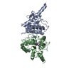
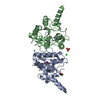
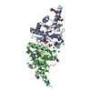
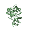
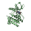
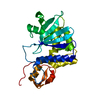
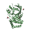
- PDB-4f78: Crystal Structure of Vancomycin Resistance D,D-dipeptidase VanXYg -
+
Open data
ID or keywords:
Loading...
-
Basic information
Entry
Database: PDB / ID: 4f78
Title
Crystal Structure of Vancomycin Resistance D,D-dipeptidase VanXYg
Components
D,D-dipeptidase/D,D-carboxypeptidase
Keywords
HYDROLASE / CENTER FOR STRUCTURAL GENOMICS OF INFECTIOUS DISEASES / CSGID / NATIONAL INSTITUTE OF ALLERGY AND INFECTIOUS DISEASES / NIAID / ALPHA+BETA PROTEIN / METALLOPEPTIDASE / HEDGEHOG/DD-PEPTIDASE FOLD / MEROPS M15B SUBFAMILY / ZN2+-DEPENDENT D / D-DIPEPTIDASE / VANCOMYCIN RESISTANCE / D-ALANINE-D-ALANINE
Resolution: 1.95→2.06 Å / Redundancy: 3.3 % / Rmerge(I) obs: 0.329 / Mean I/σ(I) obs: 3.6 / Num. unique all: 2850 / % possible all: 100
-
Processing
Software
Name
Version
Classification
HKL-3000
datacollection
PHENIX
(phenix.autosol)
modelbuilding
PHENIX
(phenix.refine: 1.7.3_928)
refinement
XDS
datareduction
SCALA
datascaling
PHENIX
phasing
Refinement
Method to determine structure: SAD / Resolution: 1.95→39.584 Å / SU ML: 0.19 / Cross valid method: THROUGHOUT / σ(F): 1.35 / Phase error: 18.5 / Stereochemistry target values: ML
Rfactor
Num. reflection
% reflection
Selection details
Rfree
0.1864
949
4.99 %
random
Rwork
0.1536
-
-
-
obs
0.1554
19734
98.35 %
-
Solvent computation
Shrinkage radii: 0.73 Å / VDW probe radii: 1 Å / Solvent model: FLAT BULK SOLVENT MODEL / Bsol: 48.654 Å2 / ksol: 0.399 e/Å3
Displacement parameters
Baniso -1
Baniso -2
Baniso -3
1-
-1.7143 Å2
0 Å2
-4.2779 Å2
2-
-
1.3494 Å2
-0 Å2
3-
-
-
0.3648 Å2
Refinement step
Cycle: LAST / Resolution: 1.95→39.584 Å
Protein
Nucleic acid
Ligand
Solvent
Total
Num. atoms
2054
0
65
187
2306
Refine LS restraints
Refine-ID
Type
Dev ideal
Number
X-RAY DIFFRACTION
f_bond_d
0.016
2201
X-RAY DIFFRACTION
f_angle_d
1.494
2966
X-RAY DIFFRACTION
f_dihedral_angle_d
14.268
840
X-RAY DIFFRACTION
f_chiral_restr
0.116
310
X-RAY DIFFRACTION
f_plane_restr
0.007
386
LS refinement shell
Resolution (Å)
Rfactor Rfree
Num. reflection Rfree
Rfactor Rwork
Num. reflection Rwork
Refine-ID
% reflection obs (%)
1.95-2.0027
0.2775
123
0.2171
2747
X-RAY DIFFRACTION
98
2.0027-2.0617
0.2068
146
0.1947
2708
X-RAY DIFFRACTION
98
2.0617-2.1282
0.2173
144
0.1755
2768
X-RAY DIFFRACTION
98
2.1282-2.2043
0.2052
137
0.1596
2715
X-RAY DIFFRACTION
98
2.2043-2.2925
0.2152
146
0.16
2811
X-RAY DIFFRACTION
98
2.2925-2.3968
0.1972
151
0.1557
2673
X-RAY DIFFRACTION
98
2.3968-2.5232
0.2262
151
0.158
2766
X-RAY DIFFRACTION
98
2.5232-2.6812
0.1956
136
0.1529
2757
X-RAY DIFFRACTION
99
2.6812-2.8882
0.1882
152
0.1515
2757
X-RAY DIFFRACTION
99
2.8882-3.1787
0.1934
153
0.1497
2758
X-RAY DIFFRACTION
99
3.1787-3.6384
0.1718
145
0.1439
2772
X-RAY DIFFRACTION
99
3.6384-4.5829
0.1564
147
0.1227
2756
X-RAY DIFFRACTION
99
4.5829-39.5926
0.1556
144
0.1596
2738
X-RAY DIFFRACTION
98
Refinement TLS params.
Method: refined / Refine-ID: X-RAY DIFFRACTION
ID
L11 (°2)
L12 (°2)
L13 (°2)
L22 (°2)
L23 (°2)
L33 (°2)
S11 (Å °)
S12 (Å °)
S13 (Å °)
S21 (Å °)
S22 (Å °)
S23 (Å °)
S31 (Å °)
S32 (Å °)
S33 (Å °)
T11 (Å2)
T12 (Å2)
T13 (Å2)
T22 (Å2)
T23 (Å2)
T33 (Å2)
Origin x (Å)
Origin y (Å)
Origin z (Å)
1
0.3823
0.0117
0.5165
0.7702
-0.0827
1.4629
-0.0166
-0.3294
-0.2666
0.1715
0.0401
0.1086
0.1831
-0.0589
-0.0657
0.1352
-0.0144
0.0118
0.1233
0.0325
0.1346
-13.4764
-13.882
-18.1936
2
1.0575
-0.424
-0.5374
1.6291
0.7564
3.0137
0.0413
-0.0201
0.0131
-0.0517
-0.0586
0.0985
0.0093
-0.1347
0.0057
0.0502
-0.0142
-0.0138
0.0542
0.005
0.1032
-17.2558
-5.3482
-31.1827
3
2.6162
-0.66
-0.0608
2.2842
-0.2919
2.1523
0.0153
-0.0502
0.1704
-0.0675
0.0561
0.0946
-0.2049
-0.1041
-0.0282
0.0715
-0.0035
0.0197
0.0411
-0.0185
0.0841
-11.8754
3.1163
-23.7725
4
2.7849
0.4257
0.4265
3.8666
-0.5094
3.098
-0.0539
-0.6402
-0.0715
0.7326
-0.0311
0.0969
0.2433
-0.0655
0.0156
0.1725
0.0405
-0.0385
0.2656
0.0223
0.0305
-3.1852
-8.8349
-5.4101
Refinement TLS group
ID
Refine-ID
Refine TLS-ID
Selection details
1
X-RAY DIFFRACTION
1
chainAandresi0:50
2
X-RAY DIFFRACTION
2
chainAandresi51:116
3
X-RAY DIFFRACTION
3
chainAandresi117:194
4
X-RAY DIFFRACTION
4
chainAandresi195:250
+
About Yorodumi
-
News
-
Feb 9, 2022. New format data for meta-information of EMDB entries
New format data for meta-information of EMDB entries
Version 3 of the EMDB header file is now the official format.
The previous official version 1.9 will be removed from the archive.
In the structure databanks used in Yorodumi, some data are registered as the other names, "COVID-19 virus" and "2019-nCoV". Here are the details of the virus and the list of structure data.
Jan 31, 2019. EMDB accession codes are about to change! (news from PDBe EMDB page)
EMDB accession codes are about to change! (news from PDBe EMDB page)
The allocation of 4 digits for EMDB accession codes will soon come to an end. Whilst these codes will remain in use, new EMDB accession codes will include an additional digit and will expand incrementally as the available range of codes is exhausted. The current 4-digit format prefixed with “EMD-” (i.e. EMD-XXXX) will advance to a 5-digit format (i.e. EMD-XXXXX), and so on. It is currently estimated that the 4-digit codes will be depleted around Spring 2019, at which point the 5-digit format will come into force.
The EM Navigator/Yorodumi systems omit the EMD- prefix.
Related info.:Q: What is EMD? / ID/Accession-code notation in Yorodumi/EM Navigator
Yorodumi is a browser for structure data from EMDB, PDB, SASBDB, etc.
This page is also the successor to EM Navigator detail page, and also detail information page/front-end page for Omokage search.
The word "yorodu" (or yorozu) is an old Japanese word meaning "ten thousand". "mi" (miru) is to see.
Related info.:EMDB / PDB / SASBDB / Comparison of 3 databanks / Yorodumi Search / Aug 31, 2016. New EM Navigator & Yorodumi / Yorodumi Papers / Jmol/JSmol / Function and homology information / Changes in new EM Navigator and Yorodumi
 Movie
Movie Controller

 Yorodumi
 Open data
 Basic information
 Components
 Keywords
 Function and homology information
 Enterococcus faecalis (bacteria)
 X-RAY DIFFRACTION /
  Authors
 Citation
 Structure visualization
 Downloads & links
 Other downloads
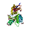


 PDBj
PDBj
 Assembly


 Mass: 65.409 Da / Num. of mol.: 1 / Source method: obtained synthetically / Formula: Zn
 Mass: 35.453 Da / Num. of mol.: 1 / Source method: obtained synthetically / Formula: Cl
 Mass: 96.063 Da / Num. of mol.: 1 / Source method: obtained synthetically / Formula: SO4
 Mass: 62.068 Da / Num. of mol.: 10 / Source method: obtained synthetically / Formula: C2H6O2
 Mass: 92.094 Da / Num. of mol.: 3 / Source method: obtained synthetically / Formula: C3H8O3
 Sample preparation
 / Beamline: 21-ID-D / Wavelength: 0.97918 Å
 Processing