 Movie
Movie Controller

[English] 日本語
 Yorodumi
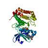
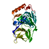
- PDB-1rl0: Crystal structure of a new ribosome-inactivating protein (RIP): d... -
+ Open data

- Basic information
| Entry | Database: PDB / ID: 1rl0 | ||||||
|---|---|---|---|---|---|---|---|
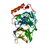
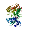
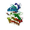
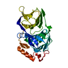
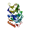
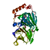
| Title | Crystal structure of a new ribosome-inactivating protein (RIP): dianthin 30 | ||||||
 Components Components | Antiviral protein DAP-30 | ||||||
 Keywords Keywords | HYDROLASE / PLANT PROTEIN / mixed beta-sheet / alpha and beta regions | ||||||
| Function / homology |  Function and homology information Function and homology informationregulation of defense response to virus / rRNA N-glycosylase / rRNA N-glycosylase activity / defense response / toxin activity / negative regulation of translation Similarity search - Function | ||||||
| Biological species |  Dianthus caryophyllus (clove pink) Dianthus caryophyllus (clove pink) | ||||||
| Method |  X-RAY DIFFRACTION / SYNCHROTRON / MOLECULAR REPLACEMENT / Resolution: 1.4 Å X-RAY DIFFRACTION / SYNCHROTRON / MOLECULAR REPLACEMENT / Resolution: 1.4 Å | ||||||
 Authors Authors | Fermani, S. / Falini, G. / Ripamonti, A. / Bolognesi, A. / Polito, L. / Stirpe, F. | ||||||
 Citation Citation | Journal: J.Struct.Biol. / Year: 2005 Title: The 1.4A structure of dianthin 30 indicates a role of surface potential at the active site of type 1 ribosome inactivating proteins Authors: Fermani, S. / Falini, G. / Ripamonti, A. / Polito, L. / Stirpe, F. / Bolognesi, A. #1: Journal: Acta Crystallogr.,Sect.D / Year: 2003Title: Crystallization and preliminary X-ray diffraction analysis of two ribosomes-inactivating proteins: lychnin and dianthin 30 Authors: Fermani, S. / Falini, G. / Ripamonti, A. / Bolognesi, A. / Polito, L. / Stirpe, F. #2: Journal: Biochim.Biophys.Acta / Year: 1993Title: Ribosome-inactivating proteins from plants Authors: Barbieri, L. / Battelli, M.G. / Stirpe, F. #3: Journal: Biochem.J. / Year: 1981Title: Dianthins, ribosome-damaging proteins with anti-viral properties from Dianthus caryophyllus L. (carnation) Authors: Stirpe, F. / Williams, D.G. / Onyon, L.G. / Legg, R.F. / Stevens, W.A. | ||||||
| History |
|
- Structure visualization
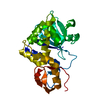
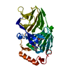
| Structure viewer | Molecule: MolmilJmol/JSmol |
|---|
- Downloads & links
-Download
| PDBx/mmCIF format | 1rl0.cif.gz | 75.1 KB | Display | PDBx/mmCIF format |
|---|---|---|---|---|
| PDB format | pdb1rl0.ent.gz | 54.6 KB | Display | PDB format |
| PDBx/mmJSON format | 1rl0.json.gz | Tree view | PDBx/mmJSON format | |
| Others |  Other downloads Other downloads |
-Validation report
| Arichive directory | https://data.pdbj.org/pub/pdb/validation_reports/rl/1rl0ftp://data.pdbj.org/pub/pdb/validation_reports/rl/1rl0 | HTTPS FTP |
|---|
-Related structure data
| Related structure data |  1qi7S S: Starting model for refinement |
|---|---|
| Similar structure data |
-Links
 PDBj
PDBj

- Assembly
| Deposited unit | 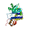
| ||||||||||
|---|---|---|---|---|---|---|---|---|---|---|---|
| 1 |
| ||||||||||
| Unit cell |
| ||||||||||
| Details | The protein is a monomeric enzyme, the asymmetric unit contains a single chain. |
-Components
| #1: Protein | Mass: 28632.631 Da / Num. of mol.: 1 / Fragment: residues 24-278 / Source method: isolated from a natural source / Source: (natural) Dianthus caryophyllus (clove pink) / Organ: leaves / References: UniProt: P24476, rRNA N-glycosylase |
|---|---|
| #2: Water | ChemComp-HOH /  Mass: 18.015 Da / Num. of mol.: 487 / Source method: isolated from a natural source / Formula: H2O Mass: 18.015 Da / Num. of mol.: 487 / Source method: isolated from a natural source / Formula: H2O |
-Experimental details
-Experiment
| Experiment | Method: X-RAY DIFFRACTION / Number of used crystals: 1 |
|---|
- Sample preparation
| Crystal | Density Matthews: 1.72 Å3/Da / Density % sol: 28 % |
|---|---|
| Crystal grow | Temperature: 293 K / pH: 4.5 Details: PEG 4000, ammonium acetate, sodium acetate, pH 4.5, VAPOR DIFFUSION, HANGING DROP, temperature 293K, pH 4.50 |
-Data collection
| Diffraction | Mean temperature: 100 K |
|---|---|
| Diffraction source | Source: SYNCHROTRON / Site: ESRF  / Beamline: ID29 / Wavelength: 1 / Beamline: ID29 / Wavelength: 1 |
| Detector | Type: ADSC QUANTUM 210 / Detector: CCD / Date: Dec 7, 2002 / Details: mirrors |
| Radiation | Monochromator: SI (111) / Protocol: SINGLE WAVELENGTH / Monochromatic (M) / Laue (L): M / Scattering type: x-ray |
| Radiation wavelength | Wavelength: 1 Å / Relative weight: 1 |
| Reflection | Resolution: 1.4→39 Å / Num. obs: 44547 / % possible obs: 93.8 % / Observed criterion σ(I): -3 / Redundancy: 6.6 % / Biso Wilson estimate: 13.1 Å2 / Rsym value: 0.063 / Net I/σ(I): 26.3 |
| Reflection shell | Resolution: 1.4→1.45 Å / Mean I/σ(I) obs: 10.4 / Rsym value: 0.255 / % possible all: 90.6 |
- Processing
| Software |
| ||||||||||||||||||||||||||||||||||||||||||||||||||||||||||||||||||||||||||||||||
|---|---|---|---|---|---|---|---|---|---|---|---|---|---|---|---|---|---|---|---|---|---|---|---|---|---|---|---|---|---|---|---|---|---|---|---|---|---|---|---|---|---|---|---|---|---|---|---|---|---|---|---|---|---|---|---|---|---|---|---|---|---|---|---|---|---|---|---|---|---|---|---|---|---|---|---|---|---|---|---|---|---|
| Refinement | Method to determine structure: MOLECULAR REPLACEMENT Starting model: PBD ENTRY 1QI7 Resolution: 1.4→36.22 Å / Rfactor Rfree error: 0.004 / Data cutoff high absF: 916080.51 / Data cutoff low absF: 0 / Isotropic thermal model: RESTRAINED / Cross valid method: THROUGHOUT / σ(F): 0 / Stereochemistry target values: ENGH & HUBER / Details: MAXIMUM LIKELIHOOD TARGET USING AMPLITUDES
| ||||||||||||||||||||||||||||||||||||||||||||||||||||||||||||||||||||||||||||||||
| Solvent computation | Solvent model: FLAT MODEL / Bsol: 78.95 Å2 / ksol: 0.33 e/Å3 | ||||||||||||||||||||||||||||||||||||||||||||||||||||||||||||||||||||||||||||||||
| Displacement parameters | Biso mean: 15.2 Å2
| ||||||||||||||||||||||||||||||||||||||||||||||||||||||||||||||||||||||||||||||||
| Refine analyze |
| ||||||||||||||||||||||||||||||||||||||||||||||||||||||||||||||||||||||||||||||||
| Refinement step | Cycle: LAST / Resolution: 1.4→36.22 Å
| ||||||||||||||||||||||||||||||||||||||||||||||||||||||||||||||||||||||||||||||||
| Refine LS restraints |
| ||||||||||||||||||||||||||||||||||||||||||||||||||||||||||||||||||||||||||||||||
| LS refinement shell | Resolution: 1.4→1.49 Å / Rfactor Rfree error: 0.015 / Total num. of bins used: 6
| ||||||||||||||||||||||||||||||||||||||||||||||||||||||||||||||||||||||||||||||||
| Xplor file |
|
