[English] 日本語
Yorodumi
- PDB-4ij8: Crystal structure of the complex of SETD8 with SAM -

+
Open data


ID or keywords:

Loading...

-
Basic information

Entry
Database: PDB / ID: 4ij8
TitleCrystal structure of the complex of SETD8 with SAM
Components
  • N-lysine methyltransferase SETD8
  • helical peptide
KeywordsTRANSFERASE / Structural Genomics Consortium / SGC / N-lysine methyltransferase
Function / homology
Function and homology information


histone H4K20 monomethyltransferase activity / lysine N-methyltransferase activity / [histone H4]-lysine20 N-methyltransferase / histone H4K20 methyltransferase activity / histone H4 methyltransferase activity / peptidyl-lysine monomethylation / polytene chromosome / protein-lysine N-methyltransferase activity / mitotic chromosome condensation / regulation of DNA damage response, signal transduction by p53 class mediator ...histone H4K20 monomethyltransferase activity / lysine N-methyltransferase activity / [histone H4]-lysine20 N-methyltransferase / histone H4K20 methyltransferase activity / histone H4 methyltransferase activity / peptidyl-lysine monomethylation / polytene chromosome / protein-lysine N-methyltransferase activity / mitotic chromosome condensation / regulation of DNA damage response, signal transduction by p53 class mediator / negative regulation of double-strand break repair via homologous recombination / histone methyltransferase activity / Transferases; Transferring one-carbon groups; Methyltransferases / Condensation of Prophase Chromosomes / regulation of signal transduction by p53 class mediator / Regulation of TP53 Activity through Methylation / PKMTs methylate histone lysines / transcription corepressor activity / cell division / negative regulation of DNA-templated transcription / chromatin / regulation of transcription by RNA polymerase II / negative regulation of transcription by RNA polymerase II / nucleoplasm / nucleus / cytosol
Similarity search - Function
Class V SAM-dependent methyltransferases / : / Histone-lysine N-methyltransferase (EC 2.1.1.43) family profile. / Beta-clip-like / SET domain / SET (Su(var)3-9, Enhancer-of-zeste, Trithorax) domain / SET domain superfamily / SET domain / SET domain profile. / SET domain ...Class V SAM-dependent methyltransferases / : / Histone-lysine N-methyltransferase (EC 2.1.1.43) family profile. / Beta-clip-like / SET domain / SET (Su(var)3-9, Enhancer-of-zeste, Trithorax) domain / SET domain superfamily / SET domain / SET domain profile. / SET domain / Beta Complex / Mainly Beta
Similarity search - Domain/homology
S-ADENOSYLMETHIONINE / N-lysine methyltransferase KMT5A
Similarity search - Component
Biological speciesHomo sapiens (human)
unidentified (others)
MethodX-RAY DIFFRACTION / SYNCHROTRON / MOLECULAR REPLACEMENT / molecular replacement / Resolution: 2 Å
AuthorsYu, W. / Tempel, W. / Li, Y. / El Bakkouri, M. / Shapira, M. / Bountra, C. / Arrowsmith, C.H. / Edwards, A.M. / Brown, P.J. / Structural Genomics Consortium (SGC)
CitationJournal: To be Published
Title: Crystal structure of the complex of SETD8 with SAM
Authors: Yu, W. / Tempel, W. / Li, Y. / El Bakkouri, M. / Shapira, M. / Bountra, C. / Arrowsmith, C.H. / Edwards, A.M. / Brown, P.J. / Structural Genomics Consortium (SGC)
History
DepositionDec 21, 2012Deposition site: RCSB / Processing site: RCSB
Revision 1.0Jan 16, 2013Provider: repository / Type: Initial release
Revision 1.1Apr 2, 2014Group: Source and taxonomy
Revision 1.2Sep 20, 2023Group: Data collection / Database references ...Data collection / Database references / Derived calculations / Refinement description
Category: chem_comp_atom / chem_comp_bond ...chem_comp_atom / chem_comp_bond / database_2 / pdbx_initial_refinement_model / struct_ref_seq_dif / struct_site
Item: _database_2.pdbx_DOI / _database_2.pdbx_database_accession ..._database_2.pdbx_DOI / _database_2.pdbx_database_accession / _struct_ref_seq_dif.details / _struct_site.pdbx_auth_asym_id / _struct_site.pdbx_auth_comp_id / _struct_site.pdbx_auth_seq_id

-
Structure visualization

Structure viewerMolecule:
MolmilJmol/JSmol

Downloads & links

-
Assembly

Deposited unit
A: N-lysine methyltransferase SETD8
B: N-lysine methyltransferase SETD8
I: helical peptide
hetero molecules


Theoretical massNumber of molelcules
Total (without water)39,10027
Polymers38,3033
Non-polymers79724
Water2,342130
1
A: N-lysine methyltransferase SETD8
B: N-lysine methyltransferase SETD8
hetero molecules


Theoretical massNumber of molelcules
Total (without water)38,23126
Polymers37,4342
Non-polymers79724
Water362
TypeNameSymmetry operationNumber
identity operation1_555x,y,z1
Buried area1830 Å2
ΔGint-10 kcal/mol
Surface area15380 Å2
MethodPISA
2
I: helical peptide


  • defined by software
  • 869 Da, 1 polymers
Theoretical massNumber of molelcules
Total (without water)8691
Polymers8691
Non-polymers00
Water0
TypeNameSymmetry operationNumber
identity operation1_555x,y,z1
MethodPISA
Unit cell
Length a, b, c (Å)101.439, 101.439, 140.799
Angle α, β, γ (deg.)90.000, 90.000, 120.000
Int Tables number178
Space group name H-MP6122
Components on special symmetry positions
IDModelComponents
11A-658-

HOH

21B-631-

HOH

-
Components

#1: Protein N-lysine methyltransferase SETD8 / H4-K20-HMTase SETD8 / Histone-lysine N-methyltransferase SETD8 / Lysine N-methyltransferase 5A / ...H4-K20-HMTase SETD8 / Histone-lysine N-methyltransferase SETD8 / Lysine N-methyltransferase 5A / PR/SET domain-containing protein 07 / PR-Set7 / PR/SET07 / SET domain-containing protein 8


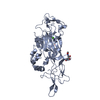
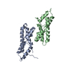
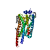
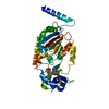
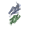
Mass: 18717.139 Da / Num. of mol.: 2 / Mutation: C343S
Source method: isolated from a genetically manipulated source
Source: (gene. exp.) Homo sapiens (human) / Gene: SETD8, KMT5A, PRSET7, SET07, SET8 / Plasmid: pHIS2 / Production host: Escherichia coli (E. coli) / Strain (production host): BL21-V2R-pRARE2
References: UniProt: Q9NQR1, Transferases; Transferring one-carbon groups; Methyltransferases, histone-lysine N-methyltransferase
#2: Protein/peptide helical peptide


Mass: 869.063 Da / Num. of mol.: 1 / Fragment: SEE REMARK 999
Source method: isolated from a genetically manipulated source
Source: (gene. exp.) unidentified (others) / Production host: Escherichia coli (E. coli)
#3: Chemical ChemComp-SAM / S-ADENOSYLMETHIONINE / S-Adenosyl methionine


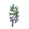
Mass: 398.437 Da / Num. of mol.: 2 / Source method: obtained synthetically / Formula: C15H22N6O5S
#4: Chemical...
ChemComp-UNX / UNKNOWN ATOM OR ION


Num. of mol.: 22 / Source method: obtained synthetically
#5: Water ChemComp-HOH / water / Water


Mass: 18.015 Da / Num. of mol.: 130 / Source method: isolated from a natural source / Formula: H2O
Sequence detailsTHE MODEL INCLUDES A DISJOINT APPARENT ALPHA-HELIX THAT COULD NOT BE ASSIGNED TO A SPECIFIC SECTION ...THE MODEL INCLUDES A DISJOINT APPARENT ALPHA-HELIX THAT COULD NOT BE ASSIGNED TO A SPECIFIC SECTION OF THE AMINO ACID SEQUENCE OF THE TARGET PROTEIN.

-
Experimental details

-
Experiment

ExperimentMethod: X-RAY DIFFRACTION / Number of used crystals: 1

-
Sample preparation

CrystalDensity Matthews: 2.73 Å3/Da / Density % sol: 54.94 %
Crystal growTemperature: 291 K / Method: vapor diffusion / pH: 7.5
Details: 1.2 M sodium citrate, 0.1 M HEPES, 10-fold excess SAM, pH 7.5, VAPOR DIFFUSION, temperature 291K

-
Data collection

DiffractionMean temperature: 100 K
Diffraction sourceSource: SYNCHROTRON / Site: CLSI / Beamline: 08ID-1 / Wavelength: 0.97949 Å
DetectorType: RAYONIX MX-300 / Detector: CCD / Date: Jan 25, 2012
RadiationMonochromator: double crystal Si(111) / Protocol: SINGLE WAVELENGTH / Monochromatic (M) / Laue (L): M / Scattering type: x-ray
Radiation wavelengthWavelength: 0.97949 Å / Relative weight: 1
ReflectionResolution: 2→87.849 Å / Num. all: 29619 / Num. obs: 29619 / % possible obs: 100 % / Redundancy: 21.6 % / Rsym value: 0.112 / Net I/σ(I): 19.7
Reflection shell

Diffraction-ID: 1

Resolution (Å)Redundancy (%)Rmerge(I) obsMean I/σ(I) obsNum. measured allNum. unique allRsym value% possible all
2-2.11220.9420.89308642310.942100
2.11-2.24220.5911.38796239900.591100
2.24-2.3921.90.4041.98301837830.404100
2.39-2.58220.2862.77772835350.286100
2.58-2.8321.90.184.27167032720.18100
2.83-3.1621.70.1136.36484129840.113100
3.16-3.6521.40.0817.95660626490.081100
3.65-4.47210.0649.24770422720.064100
4.47-6.3220.40.0619.43702118110.061100
6.32-46.92818.30.0589.22001610920.05899.7

-
Phasing

PhasingMethod: molecular replacement

-
Processing

Software
NameVersionClassificationNB
SCALA3.3.20data scaling
PHASERphasing
REFMAC5.7.0032refinement
PDB_EXTRACT3.11data extraction
XDSdata reduction
RefinementMethod to determine structure: MOLECULAR REPLACEMENT
Starting model: PDB ENTRY 1ZKK
Resolution: 2→43.96 Å / Cor.coef. Fo:Fc: 0.958 / Cor.coef. Fo:Fc free: 0.952 / WRfactor Rfree: 0.1892 / WRfactor Rwork: 0.1704 / Occupancy max: 1 / Occupancy min: 0.3 / FOM work R set: 0.8803 / SU B: 6.332 / SU ML: 0.09 / SU R Cruickshank DPI: 0.1405 / SU Rfree: 0.1248 / Cross valid method: THROUGHOUT / σ(F): 0 / ESU R: 0.14 / ESU R Free: 0.125 / Stereochemistry target values: MAXIMUM LIKELIHOOD
Details: HYDROGENS HAVE BEEN ADDED IN THE RIDING POSITIONS U VALUES : WITH TLS ADDED ARP/WARP, COOT, AND THE MOLPROBITY SERVER WERE ALSO USED DURING REFINEMENT.
RfactorNum. reflection% reflectionSelection details
Rfree0.1985 1516 5.1 %THIN SHELLS (SFTOOLS)
Rwork0.1757 ---
obs0.1769 29561 99.97 %-
Solvent computationIon probe radii: 0.8 Å / Shrinkage radii: 0.8 Å / VDW probe radii: 1.2 Å / Solvent model: MASK
Displacement parametersBiso max: 97.23 Å2 / Biso mean: 37.7619 Å2 / Biso min: 15.59 Å2
Baniso -1Baniso -2Baniso -3
1--0.14 Å2-0.14 Å2-0 Å2
2---0.14 Å2-0 Å2
3---0.44 Å2
Refinement stepCycle: LAST / Resolution: 2→43.96 Å
ProteinNucleic acidLigandSolventTotal
Num. atoms2440 0 76 130 2646
Refine LS restraints
Refine-IDTypeDev idealDev ideal targetNumber
X-RAY DIFFRACTIONr_bond_refined_d0.0150.0192575
X-RAY DIFFRACTIONr_bond_other_d0.0020.022342
X-RAY DIFFRACTIONr_angle_refined_deg1.4811.993497
X-RAY DIFFRACTIONr_angle_other_deg0.7235383
X-RAY DIFFRACTIONr_dihedral_angle_1_deg6.1445331
X-RAY DIFFRACTIONr_dihedral_angle_2_deg30.1424.107112
X-RAY DIFFRACTIONr_dihedral_angle_3_deg11.8415418
X-RAY DIFFRACTIONr_dihedral_angle_4_deg18.0861515
X-RAY DIFFRACTIONr_chiral_restr0.0860.2391
X-RAY DIFFRACTIONr_gen_planes_refined0.0050.022945
X-RAY DIFFRACTIONr_gen_planes_other0.0010.02583
X-RAY DIFFRACTIONr_mcbond_it1.6452.2241303
X-RAY DIFFRACTIONr_mcbond_other1.6452.2231302
X-RAY DIFFRACTIONr_mcangle_it2.383.3231624
LS refinement shellResolution: 2→2.052 Å / Total num. of bins used: 20
RfactorNum. reflection% reflection
Rfree0.321 2 -
Rwork0.222 2116 -
all-2118 -
obs--99.95 %
Refinement TLS params.

Method: refined / Refine-ID: X-RAY DIFFRACTION

IDL112)L122)L132)L222)L232)L332)S11 (Å °)S12 (Å °)S13 (Å °)S21 (Å °)S22 (Å °)S23 (Å °)S31 (Å °)S32 (Å °)S33 (Å °)T112)T122)T132)T222)T232)T332)Origin x (Å)Origin y (Å)Origin z (Å)
12.39180.031-0.16071.40720.4081.7877-0.07960.04870.0541-0.05580.0252-0.35960.0240.26520.05440.0592-0.01570.04470.05530.01080.1252-11.208934.09384.9505
22.8454-0.2796-0.10132.2859-0.15081.7569-0.01040.1036-0.4324-0.1415-0.14160.26940.3434-0.19250.1520.1112-0.05920.03490.0561-0.06030.1106-37.989320.17463.5645
Refinement TLS group
IDRefine-IDRefine TLS-IDAuth asym-IDAuth seq-ID
1X-RAY DIFFRACTION1A233 - 393
2X-RAY DIFFRACTION2B244 - 393

+
About Yorodumi

-
News

-
Feb 9, 2022. New format data for meta-information of EMDB entries

New format data for meta-information of EMDB entries

  • Version 3 of the EMDB header file is now the official format.
  • The previous official version 1.9 will be removed from the archive.

Related info.:EMDB header

External links:wwPDB to switch to version 3 of the EMDB data model

-
Aug 12, 2020. Covid-19 info

Covid-19 info

URL: https://pdbj.org/emnavi/covid19.php

New page: Covid-19 featured information page in EM Navigator.

Related info.:Covid-19 info / Mar 5, 2020. Novel coronavirus structure data

+
Mar 5, 2020. Novel coronavirus structure data

Novel coronavirus structure data

Related info.:Yorodumi Speices / Aug 12, 2020. Covid-19 info

External links:COVID-19 featured content - PDBj / Molecule of the Month (242):Coronavirus Proteases

+
Jan 31, 2019. EMDB accession codes are about to change! (news from PDBe EMDB page)

EMDB accession codes are about to change! (news from PDBe EMDB page)

  • The allocation of 4 digits for EMDB accession codes will soon come to an end. Whilst these codes will remain in use, new EMDB accession codes will include an additional digit and will expand incrementally as the available range of codes is exhausted. The current 4-digit format prefixed with “EMD-” (i.e. EMD-XXXX) will advance to a 5-digit format (i.e. EMD-XXXXX), and so on. It is currently estimated that the 4-digit codes will be depleted around Spring 2019, at which point the 5-digit format will come into force.
  • The EM Navigator/Yorodumi systems omit the EMD- prefix.

Related info.:Q: What is EMD? / ID/Accession-code notation in Yorodumi/EM Navigator

External links:EMDB Accession Codes are Changing Soon! / Contact to PDBj

+
Jul 12, 2017. Major update of PDB

Major update of PDB

  • wwPDB released updated PDB data conforming to the new PDBx/mmCIF dictionary.
  • This is a major update changing the version number from 4 to 5, and with Remediation, in which all the entries are updated.
  • In this update, many items about electron microscopy experimental information are reorganized (e.g. em_software).
  • Now, EM Navigator and Yorodumi are based on the updated data.

External links:wwPDB Remediation / Enriched Model Files Conforming to OneDep Data Standards Now Available in the PDB FTP Archive

-
Yorodumi

Thousand views of thousand structures

  • Yorodumi is a browser for structure data from EMDB, PDB, SASBDB, etc.
  • This page is also the successor to EM Navigator detail page, and also detail information page/front-end page for Omokage search.
  • The word "yorodu" (or yorozu) is an old Japanese word meaning "ten thousand". "mi" (miru) is to see.

Related info.:EMDB / PDB / SASBDB / Comparison of 3 databanks / Yorodumi Search / Aug 31, 2016. New EM Navigator & Yorodumi / Yorodumi Papers / Jmol/JSmol / Function and homology information / Changes in new EM Navigator and Yorodumi

Read more