 Movie
Movie Controller


+ Open data

- Basic information
| Entry | Database: PDB / ID: 3zyg | ||||||
|---|---|---|---|---|---|---|---|
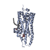
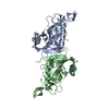
| Title | NETRING2 LAM AND EGF1 DOMAINS | ||||||
 Components Components | NETRIN-G2 | ||||||
 Keywords Keywords | CELL ADHESION / SYNAPSE | ||||||
| Function / homology |  Function and homology information Function and homology informationregulation of neuron projection arborization / postsynaptic specialization assembly / Post-translational modification: synthesis of GPI-anchored proteins / synaptic membrane adhesion / cell-cell adhesion mediator activity / regulation of neuron migration / regulation of neuron projection development / regulation of presynapse assembly / side of membrane / presynaptic active zone membrane ...regulation of neuron projection arborization / postsynaptic specialization assembly / Post-translational modification: synthesis of GPI-anchored proteins / synaptic membrane adhesion / cell-cell adhesion mediator activity / regulation of neuron migration / regulation of neuron projection development / regulation of presynapse assembly / side of membrane / presynaptic active zone membrane / axonogenesis / modulation of chemical synaptic transmission / Schaffer collateral - CA1 synapse / axon / glutamatergic synapse / extracellular region / plasma membrane Similarity search - Function | ||||||
| Biological species |  HOMO SAPIENS (human) HOMO SAPIENS (human) | ||||||
| Method |  X-RAY DIFFRACTION / SYNCHROTRON / MOLECULAR REPLACEMENT / Resolution: 2.2 Å X-RAY DIFFRACTION / SYNCHROTRON / MOLECULAR REPLACEMENT / Resolution: 2.2 Å | ||||||
 Authors Authors | Seiradake, E. / Coles, C.H. / Perestenko, P.V. / Harlos, K. / Mcilhinney, R.A.J. / Aricescu, A.R. / Jones, E.Y. | ||||||
 Citation Citation | Journal: Embo J. / Year: 2011 Title: Structural Basis for Cell Surface Patterning Through Netring-Ngl Interactions. Authors: Seiradake, E. / Coles, C.H. / Perestenko, P.V. / Harlos, K. / Mcilhinney, R.A.J. / Aricescu, A.R. / Jones, E.Y. | ||||||
| History |
|
- Structure visualization
| Structure viewer | Molecule: MolmilJmol/JSmol |
|---|
- Downloads & links
-Download
| PDBx/mmCIF format | 3zyg.cif.gz | 264.7 KB | Display | PDBx/mmCIF format |
|---|---|---|---|---|
| PDB format | pdb3zyg.ent.gz | 213.9 KB | Display | PDB format |
| PDBx/mmJSON format | 3zyg.json.gz | Tree view | PDBx/mmJSON format | |
| Others |  Other downloads Other downloads |
-Validation report
| Arichive directory | https://data.pdbj.org/pub/pdb/validation_reports/zy/3zygftp://data.pdbj.org/pub/pdb/validation_reports/zy/3zyg | HTTPS FTP |
|---|
-Related structure data
| Related structure data |  3zyiC 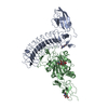 3zyjC 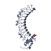 3zynC 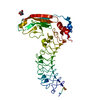 3zyoC  1tvgS C: citing same article ( S: Starting model for refinement |
|---|---|
| Similar structure data |


-Links
 PDBj
PDBj

- Assembly
| Deposited unit | 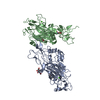
| ||||||||
|---|---|---|---|---|---|---|---|---|---|
| 1 | 
| ||||||||
| 2 | 
| ||||||||
| Unit cell |
|
-Components
| #1: Protein | Mass: 40685.191 Da / Num. of mol.: 2 / Fragment: LAM AND EGF1 DOMAINS, RESIDUES 423-767 Source method: isolated from a genetically manipulated source Source: (gene. exp.) HOMO SAPIENS (human) / Plasmid: PHLSEC / Cell line (production host): HEK 293 GNTI(-) / Production host: HOMO SAPIENS (human) / References: UniProt: Q96CW9#2: Sugar | ChemComp-NAG /   Type: D-saccharide, beta linking / Mass: 221.208 Da / Num. of mol.: 4 Type: D-saccharide, beta linking / Mass: 221.208 Da / Num. of mol.: 4Source method: isolated from a genetically manipulated source Formula: C8H15NO6 #3: Chemical |   Mass: 40.078 Da / Num. of mol.: 2 / Source method: obtained synthetically / Formula: Ca Mass: 40.078 Da / Num. of mol.: 2 / Source method: obtained synthetically / Formula: Ca#4: Water | ChemComp-HOH / |  Mass: 18.015 Da / Num. of mol.: 208 / Source method: isolated from a natural source / Formula: H2O Mass: 18.015 Da / Num. of mol.: 208 / Source method: isolated from a natural source / Formula: H2OHas protein modification | Y | Sequence details | N-TERMINAL SECRETION SIGNAL IS CLEAVED DURING PROTEIN PRODUCTION | |
|---|
-Experimental details
-Experiment
| Experiment | Method: X-RAY DIFFRACTION / Number of used crystals: 1 |
|---|
- Sample preparation
| Crystal | Density Matthews: 2.6 Å3/Da / Density % sol: 52.69 % / Description: NONE |
|---|---|
| Crystal grow | Details: 20% MME550, 0.1M NACL, 0.1M BICINE PH9 |
-Data collection
| Diffraction | Mean temperature: 100 K | |||||||||
|---|---|---|---|---|---|---|---|---|---|---|
| Diffraction source | Source: SYNCHROTRON / Site: Diamond  / Beamline: I03 / Wavelength: 1.0, 0.979400 / Beamline: I03 / Wavelength: 1.0, 0.979400 | |||||||||
| Detector | Type: ADSC CCD / Detector: CCD | |||||||||
| Radiation | Protocol: SINGLE WAVELENGTH / Monochromatic (M) / Laue (L): M / Scattering type: x-ray | |||||||||
| Radiation wavelength |
| |||||||||
| Reflection | Resolution: 2.2→29.2 Å / Num. obs: 42208 / % possible obs: 98.9 % / Observed criterion σ(I): -3 / Redundancy: 3.6 % / Rmerge(I) obs: 0.32 / Net I/σ(I): 6.4 | |||||||||
| Reflection shell | Resolution: 2.2→2.4 Å / Redundancy: 3.4 % / Rmerge(I) obs: 0.84 / Mean I/σ(I) obs: 2.35 / % possible all: 96.6 |
- Processing
| Software |
| ||||||||||||||||||||||||||||||||||||||||||||||||||||||||||||||||||||||||||||||||||||||||||||||||||||||||||||||||||||||||||||||||||||||||||||||||||||||||||||||||||||||||||||||||||||||
|---|---|---|---|---|---|---|---|---|---|---|---|---|---|---|---|---|---|---|---|---|---|---|---|---|---|---|---|---|---|---|---|---|---|---|---|---|---|---|---|---|---|---|---|---|---|---|---|---|---|---|---|---|---|---|---|---|---|---|---|---|---|---|---|---|---|---|---|---|---|---|---|---|---|---|---|---|---|---|---|---|---|---|---|---|---|---|---|---|---|---|---|---|---|---|---|---|---|---|---|---|---|---|---|---|---|---|---|---|---|---|---|---|---|---|---|---|---|---|---|---|---|---|---|---|---|---|---|---|---|---|---|---|---|---|---|---|---|---|---|---|---|---|---|---|---|---|---|---|---|---|---|---|---|---|---|---|---|---|---|---|---|---|---|---|---|---|---|---|---|---|---|---|---|---|---|---|---|---|---|---|---|---|---|
| Refinement | Method to determine structure: MOLECULAR REPLACEMENT Starting model: PDB ENTRY 1TVG Resolution: 2.2→30 Å / Cor.coef. Fo:Fc: 0.932 / Cor.coef. Fo:Fc free: 0.893 / SU B: 13.952 / SU ML: 0.181 / Cross valid method: THROUGHOUT / ESU R: 0.301 / ESU R Free: 0.245 / Stereochemistry target values: MAXIMUM LIKELIHOOD Details: HYDROGENS HAVE BEEN USED IF PRESENT PRESENT IN THE INPUT.
| ||||||||||||||||||||||||||||||||||||||||||||||||||||||||||||||||||||||||||||||||||||||||||||||||||||||||||||||||||||||||||||||||||||||||||||||||||||||||||||||||||||||||||||||||||||||
| Solvent computation | Ion probe radii: 0.8 Å / Shrinkage radii: 0.8 Å / VDW probe radii: 1.2 Å / Solvent model: MASK | ||||||||||||||||||||||||||||||||||||||||||||||||||||||||||||||||||||||||||||||||||||||||||||||||||||||||||||||||||||||||||||||||||||||||||||||||||||||||||||||||||||||||||||||||||||||
| Displacement parameters | Biso mean: 25.277 Å2
| ||||||||||||||||||||||||||||||||||||||||||||||||||||||||||||||||||||||||||||||||||||||||||||||||||||||||||||||||||||||||||||||||||||||||||||||||||||||||||||||||||||||||||||||||||||||
| Refinement step | Cycle: LAST / Resolution: 2.2→30 Å
| ||||||||||||||||||||||||||||||||||||||||||||||||||||||||||||||||||||||||||||||||||||||||||||||||||||||||||||||||||||||||||||||||||||||||||||||||||||||||||||||||||||||||||||||||||||||
| Refine LS restraints |
|