[English] 日本語
Yorodumi
- PDB-1tvg: X-ray structure of human PP25 gene product, HSPC034. Northeast St... -

+
Open data


ID or keywords:

Loading...

-
Basic information

Entry
Database: PDB / ID: 1tvg
TitleX-ray structure of human PP25 gene product, HSPC034. Northeast Structural Genomics Target HR1958.
ComponentsLOC51668 protein
KeywordsCELL CYCLE / Structural Genomics / PSI / Protein Structure Initiative / Northeast Structural Genomics Consortium / NESG
Function / homology
Function and homology information


intraciliary anterograde transport / intraciliary transport particle B / left/right axis specification / ciliary tip / Intraflagellar transport / smoothened signaling pathway / cilium assembly / lung development / kidney development / skeletal system development ...intraciliary anterograde transport / intraciliary transport particle B / left/right axis specification / ciliary tip / Intraflagellar transport / smoothened signaling pathway / cilium assembly / lung development / kidney development / skeletal system development / cilium / protein transport / heart development / spermatogenesis / cell differentiation / centrosome / metal ion binding
Similarity search - Function
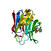
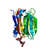
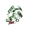
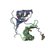
Intraflagellar transport protein 25 / F5/8 type C domain / Coagulation factor 5/8 C-terminal domain / Galactose-binding domain-like / Galactose-binding-like domain superfamily / Jelly Rolls / Sandwich / Mainly Beta
Similarity search - Domain/homology
SAMARIUM (III) ION / Intraflagellar transport protein 25 homolog
Similarity search - Component
Biological speciesHomo sapiens (human)
MethodX-RAY DIFFRACTION / SYNCHROTRON / SAD for Sm derivative / Resolution: 1.6 Å
AuthorsKuzin, A.P. / Vorobiev, S.M. / Lee, I. / Acton, T.B. / Montelione, G.T. / Tong, L. / Hunt, J.F. / Northeast Structural Genomics Consortium (NESG)
CitationJournal: Proteins / Year: 2009
Title: Improving NMR protein structure quality by Rosetta refinement: a molecular replacement study.
Authors: Ramelot, T.A. / Raman, S. / Kuzin, A.P. / Xiao, R. / Ma, L.C. / Acton, T.B. / Hunt, J.F. / Montelione, G.T. / Baker, D. / Kennedy, M.A.
History
DepositionJun 29, 2004Deposition site: RCSB / Processing site: RCSB
Revision 1.0Nov 9, 2004Provider: repository / Type: Initial release
Revision 1.1Apr 30, 2008Group: Version format compliance
Revision 1.2Jul 13, 2011Group: Version format compliance
Revision 1.3Feb 14, 2024Group: Data collection / Database references / Derived calculations
Category: chem_comp_atom / chem_comp_bond ...chem_comp_atom / chem_comp_bond / database_2 / pdbx_database_related / pdbx_struct_conn_angle / struct_conn / struct_ref_seq_dif / struct_site
Item: _database_2.pdbx_DOI / _database_2.pdbx_database_accession ..._database_2.pdbx_DOI / _database_2.pdbx_database_accession / _pdbx_database_related.db_name / _pdbx_struct_conn_angle.ptnr1_auth_comp_id / _pdbx_struct_conn_angle.ptnr1_auth_seq_id / _pdbx_struct_conn_angle.ptnr1_label_asym_id / _pdbx_struct_conn_angle.ptnr1_label_atom_id / _pdbx_struct_conn_angle.ptnr1_label_comp_id / _pdbx_struct_conn_angle.ptnr1_label_seq_id / _pdbx_struct_conn_angle.ptnr3_auth_comp_id / _pdbx_struct_conn_angle.ptnr3_auth_seq_id / _pdbx_struct_conn_angle.ptnr3_label_asym_id / _pdbx_struct_conn_angle.ptnr3_label_atom_id / _pdbx_struct_conn_angle.ptnr3_label_comp_id / _pdbx_struct_conn_angle.ptnr3_label_seq_id / _pdbx_struct_conn_angle.value / _struct_conn.pdbx_dist_value / _struct_conn.ptnr1_auth_comp_id / _struct_conn.ptnr1_auth_seq_id / _struct_conn.ptnr1_label_asym_id / _struct_conn.ptnr1_label_atom_id / _struct_conn.ptnr1_label_comp_id / _struct_conn.ptnr1_label_seq_id / _struct_conn.ptnr1_symmetry / _struct_conn.ptnr2_auth_comp_id / _struct_conn.ptnr2_auth_seq_id / _struct_conn.ptnr2_label_asym_id / _struct_conn.ptnr2_label_atom_id / _struct_conn.ptnr2_label_comp_id / _struct_conn.ptnr2_label_seq_id / _struct_conn.ptnr2_symmetry / _struct_ref_seq_dif.details / _struct_site.pdbx_auth_asym_id / _struct_site.pdbx_auth_comp_id / _struct_site.pdbx_auth_seq_id

-
Structure visualization

Structure viewerMolecule:
MolmilJmol/JSmol

Downloads & links

-
Assembly

Deposited unit
A: LOC51668 protein
hetero molecules


Theoretical massNumber of molelcules
Total (without water)17,6333
Polymers17,4431
Non-polymers1902
Water2,090116
1


  • Idetical with deposited unit
  • defined by author
TypeNameSymmetry operationNumber
identity operation1_555x,y,z1
Unit cell
Length a, b, c (Å)70.974, 41.617, 46.779
Angle α, β, γ (deg.)90.00, 102.19, 90.00
Int Tables number5
Space group name H-MC121

-
Components

#1: Protein LOC51668 protein / Anaphase-Promoting Complex / subunit 10 (APC10) / HR1958


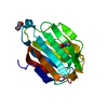
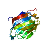
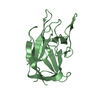
Mass: 17442.545 Da / Num. of mol.: 1
Source method: isolated from a genetically manipulated source
Details: This protein is probably a mutant because ASP109 is deleted.
Source: (gene. exp.) Homo sapiens (human) / References: UniProt: Q9Y547
#2: Chemical ChemComp-CA / CALCIUM ION


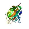
Mass: 40.078 Da / Num. of mol.: 1 / Source method: obtained synthetically / Formula: Ca
#3: Chemical ChemComp-SM / SAMARIUM (III) ION


Mass: 150.360 Da / Num. of mol.: 1 / Source method: obtained synthetically / Formula: Sm
#4: Water ChemComp-HOH / water


Mass: 18.015 Da / Num. of mol.: 116 / Source method: isolated from a natural source / Formula: H2O

-
Experimental details

-
Experiment

ExperimentMethod: X-RAY DIFFRACTION / Number of used crystals: 1

-
Sample preparation

CrystalDensity Matthews: 2 Å3/Da / Density % sol: 39 %
Crystal growTemperature: 293 K / Method: vapor diffusion, hanging drop
Details: 18% PEG, 200 mM CaCl(2), VAPOR DIFFUSION, HANGING DROP, temperature 293K

-
Data collection

DiffractionMean temperature: 100 K
Diffraction sourceSource: SYNCHROTRON / Site: NSLS / Beamline: X4A / Wavelength: 0.97896 Å
DetectorType: ADSC QUANTUM 4 / Detector: CCD / Date: Apr 2, 2004
RadiationProtocol: SINGLE WAVELENGTH / Monochromatic (M) / Laue (L): M / Scattering type: x-ray
Radiation wavelengthWavelength: 0.97896 Å / Relative weight: 1
ReflectionResolution: 1.4→30 Å / Num. obs: 48903 / Observed criterion σ(I): -3 / Redundancy: 3.59 % / Biso Wilson estimate: 11.2 Å2 / Rsym value: 0.071 / Net I/σ(I): 18.47
Reflection shellResolution: 1.4→1.45 Å / Rmerge(I) obs: 0.407 / Mean I/σ(I) obs: 1.65 / % possible all: 60

-
Processing

Software
NameVersionClassification
CNS1.1refinement
DENZOdata reduction
SCALEPACKdata scaling
SOLVEphasing
RefinementMethod to determine structure: SAD for Sm derivative / Resolution: 1.6→29.74 Å / Rfactor Rfree error: 0.004 / Data cutoff high absF: 488056.06 / Data cutoff low absF: 0 / Isotropic thermal model: RESTRAINED / Cross valid method: THROUGHOUT / σ(F): 1
RfactorNum. reflection% reflectionSelection details
Rfree0.244 3200 9.9 %RANDOM
Rwork0.215 ---
obs0.215 32294 93.5 %-
all-34479 --
Solvent computationSolvent model: FLAT MODEL / Bsol: 38.6769 Å2 / ksol: 0.382308 e/Å3
Displacement parametersBiso mean: 14.7 Å2
Baniso -1Baniso -2Baniso -3
1--1.42 Å20 Å2-0.23 Å2
2--0.92 Å20 Å2
3---0.5 Å2
Refine analyze
FreeObs
Luzzati coordinate error0.23 Å0.19 Å
Luzzati d res low-5 Å
Luzzati sigma a0.06 Å0.06 Å
Refinement stepCycle: LAST / Resolution: 1.6→29.74 Å
ProteinNucleic acidLigandSolventTotal
Num. atoms1084 0 2 116 1202
Refine LS restraints
Refine-IDTypeDev idealDev ideal target
X-RAY DIFFRACTIONc_bond_d0.005
X-RAY DIFFRACTIONc_bond_d_na
X-RAY DIFFRACTIONc_bond_d_prot
X-RAY DIFFRACTIONc_angle_d
X-RAY DIFFRACTIONc_angle_d_na
X-RAY DIFFRACTIONc_angle_d_prot
X-RAY DIFFRACTIONc_angle_deg1.3
X-RAY DIFFRACTIONc_angle_deg_na
X-RAY DIFFRACTIONc_angle_deg_prot
X-RAY DIFFRACTIONc_dihedral_angle_d26.5
X-RAY DIFFRACTIONc_dihedral_angle_d_na
X-RAY DIFFRACTIONc_dihedral_angle_d_prot
X-RAY DIFFRACTIONc_improper_angle_d0.66
X-RAY DIFFRACTIONc_improper_angle_d_na
X-RAY DIFFRACTIONc_improper_angle_d_prot
X-RAY DIFFRACTIONc_mcbond_it1.341.5
X-RAY DIFFRACTIONc_mcangle_it1.982
X-RAY DIFFRACTIONc_scbond_it2.252
X-RAY DIFFRACTIONc_scangle_it3.332.5
LS refinement shellResolution: 1.6→1.7 Å / Rfactor Rfree error: 0.012 / Total num. of bins used: 6
RfactorNum. reflection% reflection
Rfree0.28 501 10.3 %
Rwork0.227 4364 -
obs--84.5 %
Xplor file
Refine-IDSerial noParam fileTopol file
X-RAY DIFFRACTION1PROTEIN_REP.PARAMPROTEIN.TOP
X-RAY DIFFRACTION2WATER_REP.PARAMWATER.TOP
X-RAY DIFFRACTION3ION.PARAMION.TOP
X-RAY DIFFRACTION4ION_SM.PARAMION_SM.TOP

+
About Yorodumi

-
News

-
Feb 9, 2022. New format data for meta-information of EMDB entries

New format data for meta-information of EMDB entries

  • Version 3 of the EMDB header file is now the official format.
  • The previous official version 1.9 will be removed from the archive.

Related info.:EMDB header

External links:wwPDB to switch to version 3 of the EMDB data model

-
Aug 12, 2020. Covid-19 info

Covid-19 info

URL: https://pdbjlvh1.pdbj.org/emnavi/covid19.php

New page: Covid-19 featured information page in EM Navigator.

Related info.:Covid-19 info / Mar 5, 2020. Novel coronavirus structure data

+
Mar 5, 2020. Novel coronavirus structure data

Novel coronavirus structure data

Related info.:Yorodumi Speices / Aug 12, 2020. Covid-19 info

External links:COVID-19 featured content - PDBj / Molecule of the Month (242):Coronavirus Proteases

+
Jan 31, 2019. EMDB accession codes are about to change! (news from PDBe EMDB page)

EMDB accession codes are about to change! (news from PDBe EMDB page)

  • The allocation of 4 digits for EMDB accession codes will soon come to an end. Whilst these codes will remain in use, new EMDB accession codes will include an additional digit and will expand incrementally as the available range of codes is exhausted. The current 4-digit format prefixed with “EMD-” (i.e. EMD-XXXX) will advance to a 5-digit format (i.e. EMD-XXXXX), and so on. It is currently estimated that the 4-digit codes will be depleted around Spring 2019, at which point the 5-digit format will come into force.
  • The EM Navigator/Yorodumi systems omit the EMD- prefix.

Related info.:Q: What is EMD? / ID/Accession-code notation in Yorodumi/EM Navigator

External links:EMDB Accession Codes are Changing Soon! / Contact to PDBj

+
Jul 12, 2017. Major update of PDB

Major update of PDB

  • wwPDB released updated PDB data conforming to the new PDBx/mmCIF dictionary.
  • This is a major update changing the version number from 4 to 5, and with Remediation, in which all the entries are updated.
  • In this update, many items about electron microscopy experimental information are reorganized (e.g. em_software).
  • Now, EM Navigator and Yorodumi are based on the updated data.

External links:wwPDB Remediation / Enriched Model Files Conforming to OneDep Data Standards Now Available in the PDB FTP Archive

-
Yorodumi

Thousand views of thousand structures

  • Yorodumi is a browser for structure data from EMDB, PDB, SASBDB, etc.
  • This page is also the successor to EM Navigator detail page, and also detail information page/front-end page for Omokage search.
  • The word "yorodu" (or yorozu) is an old Japanese word meaning "ten thousand". "mi" (miru) is to see.

Related info.:EMDB / PDB / SASBDB / Comparison of 3 databanks / Yorodumi Search / Aug 31, 2016. New EM Navigator & Yorodumi / Yorodumi Papers / Jmol/JSmol / Function and homology information / Changes in new EM Navigator and Yorodumi

Read more