 Movie
Movie Controller


+ Open data

- Basic information
| Entry | Database: PDB / ID: 1g86 | ||||||
|---|---|---|---|---|---|---|---|
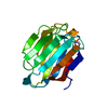
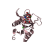
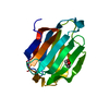
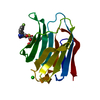
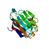
| Title | CHARCOT-LEYDEN CRYSTAL PROTEIN/N-ETHYLMALEIMIDE COMPLEX | ||||||
 Components Components | CHARCOT-LEYDEN CRYSTAL PROTEIN | ||||||
 Keywords Keywords | HYDROLASE / beta barrel | ||||||
| Function / homology |  Function and homology information Function and homology informationregulation of activated T cell proliferation / regulation of T cell cytokine production / T cell apoptotic process / regulation of T cell anergy / carbohydrate binding / extracellular matrix / identical protein binding / cytosol Similarity search - Function | ||||||
| Biological species |  Homo sapiens (human) Homo sapiens (human) | ||||||
| Method |  X-RAY DIFFRACTION / SYNCHROTRON / MOLECULAR REPLACEMENT / Resolution: 1.8 Å X-RAY DIFFRACTION / SYNCHROTRON / MOLECULAR REPLACEMENT / Resolution: 1.8 Å | ||||||
 Authors Authors | Ackerman, S.J. / Liu, L. / Kwatia, M.A. / Savage, M.P. / Leonidas, D.D. / Swaminathan, G.J. / Acharya, K.R. | ||||||
 Citation Citation | Journal: J.Biol.Chem. / Year: 2002 Title: Charcot-Leyden crystal protein (galectin-10) is not a dual function galectin with lysophospholipase activity but binds a lysophospholipase inhibitor in a novel structural fashion. Authors: Ackerman, S.J. / Liu, L. / Kwatia, M.A. / Savage, M.P. / Leonidas, D.D. / Swaminathan, G.J. / Acharya, K.R. #1: Journal: Biochemistry / Year: 1999Title: Selective Recognition of Mannose by Human Eosinophil Charcot-Leyden Crystal Protein (Galectin-10): a Crystallographic Study at 1.8 A Resolution. Authors: Swaminathan, G.J. / Leonidas, D.D. / Savage, M.P. / Ackerman, S.J. / Acharya, K.R. #2: Journal: Structure / Year: 1995Title: Crystal Structure of Human Charcot-Leyden Crystal Protein, an Eosinophil Lysophospholipase, Identifies it as a New Member of the Carbohydrate-binding Family of Galectins. Authors: Leonidas, D.D. / Elbert, B.L. / Zhou, Z. / Leffler, H. / Ackerman, S.J. / Acharya, K.R. | ||||||
| History |
|
- Structure visualization
| Structure viewer | Molecule: MolmilJmol/JSmol |
|---|
- Downloads & links
-Download
| PDBx/mmCIF format | 1g86.cif.gz | 44.7 KB | Display | PDBx/mmCIF format |
|---|---|---|---|---|
| PDB format | pdb1g86.ent.gz | 31.2 KB | Display | PDB format |
| PDBx/mmJSON format | 1g86.json.gz | Tree view | PDBx/mmJSON format | |
| Others |  Other downloads Other downloads |
-Validation report
| Arichive directory | https://data.pdbj.org/pub/pdb/validation_reports/g8/1g86ftp://data.pdbj.org/pub/pdb/validation_reports/g8/1g86 | HTTPS FTP |
|---|
-Related structure data
| Related structure data | 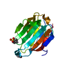 1hdkC  1lclS S: Starting model for refinement C: citing same article ( |
|---|---|
| Similar structure data |


-Links
 PDBj
PDBj- Assembly
| Deposited unit | 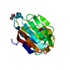
| |||||||||
|---|---|---|---|---|---|---|---|---|---|---|
| 1 |
| |||||||||
| Unit cell |
| |||||||||
| Components on special symmetry positions |
|
-Components
| #1: Protein | Mass: 16499.887 Da / Num. of mol.: 1 / Source method: isolated from a natural source / Details: EOSINOPHIL PRIMARY GRANULE / Source: (natural) Homo sapiens (human) / References: UniProt: Q05315, lysophospholipase | ||
|---|---|---|---|
| #2: Chemical |   Mass: 125.125 Da / Num. of mol.: 2 / Source method: obtained synthetically / Formula: C6H7NO2 Mass: 125.125 Da / Num. of mol.: 2 / Source method: obtained synthetically / Formula: C6H7NO2#3: Water | ChemComp-HOH / |  Mass: 18.015 Da / Num. of mol.: 82 / Source method: isolated from a natural source / Formula: H2O Mass: 18.015 Da / Num. of mol.: 82 / Source method: isolated from a natural source / Formula: H2O |
-Experimental details
-Experiment
| Experiment | Method: X-RAY DIFFRACTION / Number of used crystals: 2 |
|---|
- Sample preparation
| Crystal | Density Matthews: 2.86 Å3/Da / Density % sol: 57.01 % |
|---|---|
| Crystal grow | Temperature: 293 K / Method: vapor diffusion, hanging drop / pH: 7 Details: Tris-Acetate 0.1M, pH7.0, VAPOR DIFFUSION, HANGING DROP, temperature 293K |
-Data collection
| Diffraction |
| ||||||||||||||||||
|---|---|---|---|---|---|---|---|---|---|---|---|---|---|---|---|---|---|---|---|
| Diffraction source |
| ||||||||||||||||||
| Detector |
| ||||||||||||||||||
| Radiation |
| ||||||||||||||||||
| Radiation wavelength |
| ||||||||||||||||||
| Reflection | Resolution: 1.8→20 Å / Num. all: 17870 / Num. obs: 17573 / % possible obs: 93.3 % / Observed criterion σ(F): 0 / Observed criterion σ(I): 0 / Redundancy: 4.08 % / Biso Wilson estimate: 15.5 Å2 / Rmerge(I) obs: 0.132 / Rsym value: 0.053 / Net I/σ(I): 12.8 | ||||||||||||||||||
| Reflection shell | Resolution: 1.8→1.91 Å / Redundancy: 5.2 % / Rmerge(I) obs: 0.233 / Mean I/σ(I) obs: 7.2 / Num. unique all: 2832 / Rsym value: 0.182 / % possible all: 91.6 |

- Processing
| Software |
| ||||||||||||||||||||||||||||||||||||
|---|---|---|---|---|---|---|---|---|---|---|---|---|---|---|---|---|---|---|---|---|---|---|---|---|---|---|---|---|---|---|---|---|---|---|---|---|---|
| Refinement | Method to determine structure: MOLECULAR REPLACEMENT Starting model: 1LCL Resolution: 1.8→20 Å / Rfactor Rfree error: 0.008 / Data cutoff high absF: 10000000 / Data cutoff low absF: 0 / Isotropic thermal model: RESTRAINED / Cross valid method: THROUGHOUT / σ(F): 0 / σ(I): 0 / Stereochemistry target values: Engh & Huber
| ||||||||||||||||||||||||||||||||||||
| Displacement parameters | Biso mean: 24.1 Å2
| ||||||||||||||||||||||||||||||||||||
| Refine analyze |
| ||||||||||||||||||||||||||||||||||||
| Refinement step | Cycle: LAST / Resolution: 1.8→20 Å
| ||||||||||||||||||||||||||||||||||||
| Refine LS restraints |
| ||||||||||||||||||||||||||||||||||||
| LS refinement shell | Resolution: 1.8→1.91 Å / Rfactor Rfree error: 0.029 / Total num. of bins used: 6
| ||||||||||||||||||||||||||||||||||||
| Xplor file |
|
