 Movie
Movie Controller


+ Open data

- Basic information
| Entry | Database: PDB / ID: 1hdk | |||||||||
|---|---|---|---|---|---|---|---|---|---|---|
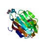
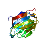
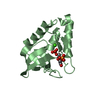
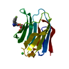
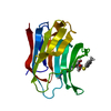
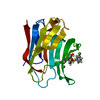
| Title | Charcot-Leyden Crystal Protein - pCMBS Complex | |||||||||
 Components Components | EOSINOPHIL LYSOPHOSPHOLIPASE | |||||||||
 Keywords Keywords | SERINE ESTERASE / GALECTIN-10 / EOSINOPHIL LYSOPHOSPHOLIPASE | |||||||||
| Function / homology |  Function and homology information Function and homology informationregulation of activated T cell proliferation / regulation of T cell cytokine production / T cell apoptotic process / regulation of T cell anergy / carbohydrate binding / extracellular matrix / identical protein binding / cytosol Similarity search - Function | |||||||||
| Biological species |  HOMO SAPIENS (human) HOMO SAPIENS (human) | |||||||||
| Method |  X-RAY DIFFRACTION / SYNCHROTRON / MIR / Resolution: 1.8 Å X-RAY DIFFRACTION / SYNCHROTRON / MIR / Resolution: 1.8 Å | |||||||||
 Authors Authors | Ackerman, S.J. / Savage, M.P. / Liu, L. / Leonidas, D.D. / Kwatia, M.A. / Swaminathan, G.J. / Acharya, K.R. | |||||||||
 Citation Citation | Journal: J.Biol.Chem. / Year: 2002 Title: Charcot-Leyden Crystal Protein (Galectin-10) is not a Dual Function Galectin with Lysophospholipase Activity But Binds a Lysophospholipase Inhibitor in a Novel Structural Fashion. Authors: Ackerman, S.J. / Liu, L. / Kwatia, M.A. / Savage, M.P. / Leonidas, D.D. / Swaminathan, G.J. / Acharya, K.R. #1: Journal: Biochemistry / Year: 1999Title: Selective Recognition of Mannose by Human Eosinophil Charcot-Leyden Crystal Protein (Galectin-10): A Crystallographic Study at 1.8 A Resolution Authors: Swaminathan, G.J. / Leonidas, D.D. / Savage, M.P. / Ackerman, S.J. / Acharya, K.R. #2: Journal: Structure / Year: 1995Title: Crystal Structure of Human Charcot-Leyden Crystal Protein, an Eosinophil Lysophospholipase Identifies It as a New Member of the Carbohydrate-Binding Family of Galectins Authors: Leonidas, D.D. / Elbert, B.L. / Zhou, Z. / Leffler, H. / Ackerman, S.J. / Acharya, K.R. | |||||||||
| History |
|
- Structure visualization
| Structure viewer | Molecule: MolmilJmol/JSmol |
|---|
- Downloads & links
-Download
| PDBx/mmCIF format | 1hdk.cif.gz | 44.9 KB | Display | PDBx/mmCIF format |
|---|---|---|---|---|
| PDB format | pdb1hdk.ent.gz | 31.5 KB | Display | PDB format |
| PDBx/mmJSON format | 1hdk.json.gz | Tree view | PDBx/mmJSON format | |
| Others |  Other downloads Other downloads |
-Validation report
| Arichive directory | https://data.pdbj.org/pub/pdb/validation_reports/hd/1hdkftp://data.pdbj.org/pub/pdb/validation_reports/hd/1hdk | HTTPS FTP |
|---|
-Related structure data


-Links
 PDBj
PDBj- Assembly
| Deposited unit | 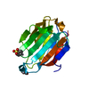
| |||||||||
|---|---|---|---|---|---|---|---|---|---|---|
| 1 |
| |||||||||
| Unit cell |
| |||||||||
| Components on special symmetry positions |
| |||||||||
| Details | BIOLOGICAL_UNIT: MONOMERA CRYSTAL PACKING DIMERIC ASSEMBLY CAN BE GENERATED USINGTHE SYMMETRY OPERATION -X, Y, 1/2 -Z. THIS CASE OF STRONGCRYSTAL PACKING HAS A DIFFERENCE IN ACCESSIBLE SURFACE AREAPER CHAIN BETWEEN THE ISOLATED CHAIN AND THAT FOR THE CHAININ THE COMPLEX OF 721.9 ANGSTROM**2 |
-Components
| #1: Protein | Mass: 16368.690 Da / Num. of mol.: 1 / Source method: isolated from a natural source / Details: COVALENTLY BOUND TO PCMBS / Source: (natural) HOMO SAPIENS (human) / Cell: EOSINOPHIL / Cellular location: PRIMARY GRANULE / Organelle: GRANULE / Tissue: BLOOD / References: UniProt: Q05315, lysophospholipase | ||||
|---|---|---|---|---|---|
| #2: Chemical |   Mass: 357.757 Da / Num. of mol.: 2 / Source method: obtained synthetically / Formula: C6H5HgO3S Mass: 357.757 Da / Num. of mol.: 2 / Source method: obtained synthetically / Formula: C6H5HgO3S#3: Water | ChemComp-HOH / |  Mass: 18.015 Da / Num. of mol.: 80 / Source method: isolated from a natural source / Formula: H2O Mass: 18.015 Da / Num. of mol.: 80 / Source method: isolated from a natural source / Formula: H2OCompound details | FUNCTION: MAY HAVE BOTH, LYSOPHOSPHOLIPASE AND CARBOHYDRATE- BINDING ACTIVITIES. CATALYTIC ACTIVITY: ...FUNCTION: MAY HAVE BOTH, LYSOPHOSPH | |
-Experimental details
-Experiment
| Experiment | Method: X-RAY DIFFRACTION / Number of used crystals: 2 |
|---|
- Sample preparation
| Crystal | Density Matthews: 2.81 Å3/Da / Density % sol: 55.87 % | ||||||||||||||||||||
|---|---|---|---|---|---|---|---|---|---|---|---|---|---|---|---|---|---|---|---|---|---|
| Crystal grow | Method: vapor diffusion, hanging drop / pH: 7 Details: TRIS-ACETATE PH7.0 100MM HANGING DROP VAPOR DIFFUSION, pH 7.00 | ||||||||||||||||||||
| Crystal grow | *PLUS Temperature: 16 ℃ / pH: 10.5 / Method: vapor diffusion, hanging dropDetails: Leonidas, D.D., (1995) Structure (London), 3, 1379. | ||||||||||||||||||||
| Components of the solutions | *PLUS
|
-Data collection
| Diffraction | Mean temperature: 293 K |
|---|---|
| Diffraction source | Source: SYNCHROTRON / Site: EMBL/DESY, HAMBURG  / Beamline: X31 / Wavelength: 0.92 / Beamline: X31 / Wavelength: 0.92 |
| Detector | Type: MARRESEARCH / Detector: IMAGE PLATE / Date: Feb 15, 1996 / Details: MIRRORS |
| Radiation | Protocol: SINGLE WAVELENGTH / Monochromatic (M) / Laue (L): M / Scattering type: x-ray |
| Radiation wavelength | Wavelength: 0.92 Å / Relative weight: 1 |
| Reflection | Resolution: 1.8→20 Å / Num. obs: 16423 / % possible obs: 87.8 % / Observed criterion σ(I): 0 / Redundancy: 1.5 % / Biso Wilson estimate: 17.9 Å2 / Rmerge(I) obs: 0.079 / Net I/σ(I): 12.7 |
| Reflection shell | Resolution: 1.8→1.87 Å / Redundancy: 1.4 % / Rmerge(I) obs: 0.127 / Mean I/σ(I) obs: 3.4 / % possible all: 72.4 |
| Reflection | *PLUS Lowest resolution: 99 Å / Num. obs: 16447 / % possible obs: 87.7 % / Num. measured all: 105494 |
- Processing
| Software |
| ||||||||||||||||||||||||||||||||||||||||||||||||||||||||||||||||||||||||||||||||
|---|---|---|---|---|---|---|---|---|---|---|---|---|---|---|---|---|---|---|---|---|---|---|---|---|---|---|---|---|---|---|---|---|---|---|---|---|---|---|---|---|---|---|---|---|---|---|---|---|---|---|---|---|---|---|---|---|---|---|---|---|---|---|---|---|---|---|---|---|---|---|---|---|---|---|---|---|---|---|---|---|---|
| Refinement | Method to determine structure: MIR / Resolution: 1.8→20 Å / Rfactor Rfree error: 0.008 / Data cutoff high absF: 10000000 / Data cutoff low absF: 0.001 / Isotropic thermal model: RESTRAINED / Cross valid method: THROUGHOUT / σ(F): 0 Details: BULK SOLVENT MODEL USED THE LAST TWO RESIDUES WERE NOT VISIBLE IN THE ELECTRON DENSITY MAPS
| ||||||||||||||||||||||||||||||||||||||||||||||||||||||||||||||||||||||||||||||||
| Displacement parameters | Biso mean: 21 Å2
| ||||||||||||||||||||||||||||||||||||||||||||||||||||||||||||||||||||||||||||||||
| Refine analyze |
| ||||||||||||||||||||||||||||||||||||||||||||||||||||||||||||||||||||||||||||||||
| Refinement step | Cycle: LAST / Resolution: 1.8→20 Å
| ||||||||||||||||||||||||||||||||||||||||||||||||||||||||||||||||||||||||||||||||
| Refine LS restraints |
| ||||||||||||||||||||||||||||||||||||||||||||||||||||||||||||||||||||||||||||||||
| LS refinement shell | Resolution: 1.8→1.91 Å / Rfactor Rfree error: 0.03 / Total num. of bins used: 6
| ||||||||||||||||||||||||||||||||||||||||||||||||||||||||||||||||||||||||||||||||
| Xplor file |
| ||||||||||||||||||||||||||||||||||||||||||||||||||||||||||||||||||||||||||||||||
| Software | *PLUS Name: X-PLOR / Version: 3.851 / Classification: refinement | ||||||||||||||||||||||||||||||||||||||||||||||||||||||||||||||||||||||||||||||||
| Refine LS restraints | *PLUS
|
