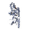
TRANSPORT PROTEIN / ION TRANSPORT-COMPLEX / CALCIUM CHANNEL BETA SUBUNIT / AID DOAMIN / ION TRANSPORT / IONIC CHANNEL / VOLTAGE-GATED CHANNEL / SH3 DOMAIN
Function / homology
Function and homology information
voltage-gated calcium channel activity involved in regulation of presynaptic cytosolic calcium levels / gamma-aminobutyric acid secretion / Presynaptic depolarization and calcium channel opening / positive regulation of protein localization to nucleolus / high voltage-gated calcium channel activity / cAMP metabolic process / detection of light stimulus involved in visual perception / Peyer's patch development / muscle cell development / neuronal action potential propagation ...voltage-gated calcium channel activity involved in regulation of presynaptic cytosolic calcium levels / gamma-aminobutyric acid secretion / Presynaptic depolarization and calcium channel opening / positive regulation of protein localization to nucleolus / high voltage-gated calcium channel activity / cAMP metabolic process / detection of light stimulus involved in visual perception / Peyer's patch development / muscle cell development / neuronal action potential propagation / nervous system process / adult walking behavior / voltage-gated calcium channel complex / negative regulation of G1/S transition of mitotic cell cycle / gamma-aminobutyric acid signaling pathway / regulation of synaptic vesicle exocytosis / neuromuscular junction development / voltage-gated calcium channel activity / spleen development / thymus development / cellular response to leukemia inhibitory factor / calcium channel regulator activity / synaptic transmission, glutamatergic / regulation of membrane potential / calcium ion transmembrane transport / calcium ion transport / T cell receptor signaling pathway / presynapse / chemical synaptic transmission / nuclear speck / negative regulation of cell population proliferation / synapse / protein kinase binding / glutamatergic synapse / plasma membrane Similarity search - Function
SHEET THE SHEET STRUCTURE OF THIS MOLECULE IS BIFURCATED. IN ORDER TO REPRESENT THIS FEATURE IN ... SHEET THE SHEET STRUCTURE OF THIS MOLECULE IS BIFURCATED. IN ORDER TO REPRESENT THIS FEATURE IN THE SHEET RECORDS BELOW, TWO SHEETS ARE DEFINED.
In the structure databanks used in Yorodumi, some data are registered as the other names, "COVID-19 virus" and "2019-nCoV". Here are the details of the virus and the list of structure data.
Jan 31, 2019. EMDB accession codes are about to change! (news from PDBe EMDB page)
EMDB accession codes are about to change! (news from PDBe EMDB page)
The allocation of 4 digits for EMDB accession codes will soon come to an end. Whilst these codes will remain in use, new EMDB accession codes will include an additional digit and will expand incrementally as the available range of codes is exhausted. The current 4-digit format prefixed with “EMD-” (i.e. EMD-XXXX) will advance to a 5-digit format (i.e. EMD-XXXXX), and so on. It is currently estimated that the 4-digit codes will be depleted around Spring 2019, at which point the 5-digit format will come into force.
The EM Navigator/Yorodumi systems omit the EMD- prefix.
Related info.:Q: What is EMD? / ID/Accession-code notation in Yorodumi/EM Navigator
Yorodumi is a browser for structure data from EMDB, PDB, SASBDB, etc.
This page is also the successor to EM Navigator detail page, and also detail information page/front-end page for Omokage search.
The word "yorodu" (or yorozu) is an old Japanese word meaning "ten thousand". "mi" (miru) is to see.
Related info.:EMDB / PDB / SASBDB / Comparison of 3 databanks / Yorodumi Search / Aug 31, 2016. New EM Navigator & Yorodumi / Yorodumi Papers / Jmol/JSmol / Function and homology information / Changes in new EM Navigator and Yorodumi
 Movie
Movie Controller


 Open data
 Basic information
 Components
 Keywords
 Function and homology information
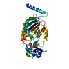
 X-RAY DIFFRACTION /
  Authors
 Citation
 Structure visualization
 Downloads & links
 Other downloads
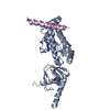


 PDBj
PDBj Assembly

 Sample preparation
 / Beamline: X4A / Wavelength: 0.9791
  Processing