 Movie
Movie Controller

[English] 日本語
 Yorodumi
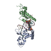
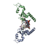
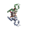
- PDB-5cq9: Crystal structure of SopD2, a type III secreted virulence effecto... -
+ Open data

- Basic information
| Entry | Database: PDB / ID: 5cq9 | ||||||
|---|---|---|---|---|---|---|---|
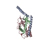
| Title | Crystal structure of SopD2, a type III secreted virulence effector from Salmonella enterica | ||||||
 Components Components |
| ||||||
 Keywords Keywords | CELL INVASION / T3SS effector protein / Structural Genomics / Montreal-Kingston Bacterial Structural Genomics Initiative / BSGI | ||||||
| Function / homology | Salmonella outer protein D / Salmonella outer protein D / protein secretion by the type III secretion system / host cell membrane / host cell plasma membrane / extracellular region / membrane / Secreted effector protein sopD2 Function and homology information Function and homology information | ||||||
| Biological species |  Salmonella typhimurium (bacteria)Salmonella enterica subsp. enterica serovar Typhimurium (bacteria) Salmonella typhimurium (bacteria)Salmonella enterica subsp. enterica serovar Typhimurium (bacteria) | ||||||
| Method |  X-RAY DIFFRACTION / SYNCHROTRON / MOLECULAR REPLACEMENT / Resolution: 3 Å X-RAY DIFFRACTION / SYNCHROTRON / MOLECULAR REPLACEMENT / Resolution: 3 Å | ||||||
 Authors Authors | Shi, R. / Cygler, M. / Montreal-Kingston Bacterial Structural Genomics Initiative (BSGI) | ||||||
| Funding support |  Canada, 1items Canada, 1items
| ||||||
 Citation Citation | Journal: Cell Rep / Year: 2015 Title: Salmonella Disrupts Host Endocytic Trafficking by SopD2-Mediated Inhibition of Rab7. Authors: D'Costa, V.M. / Braun, V. / Landekic, M. / Shi, R. / Proteau, A. / McDonald, L. / Cygler, M. / Grinstein, S. / Brumell, J.H. | ||||||
| History |
|
- Structure visualization
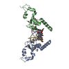
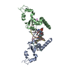
| Structure viewer | Molecule: MolmilJmol/JSmol |
|---|
- Downloads & links
-Download
| PDBx/mmCIF format | 5cq9.cif.gz | 239.3 KB | Display | PDBx/mmCIF format |
|---|---|---|---|---|
| PDB format | pdb5cq9.ent.gz | 196.9 KB | Display | PDB format |
| PDBx/mmJSON format | 5cq9.json.gz | Tree view | PDBx/mmJSON format | |
| Others |  Other downloads Other downloads |
-Validation report
| Arichive directory | https://data.pdbj.org/pub/pdb/validation_reports/cq/5cq9ftp://data.pdbj.org/pub/pdb/validation_reports/cq/5cq9 | HTTPS FTP |
|---|
-Related structure data
| Related structure data | 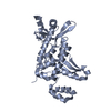 5cpcSC S: Starting model for refinement C: citing same article ( |
|---|---|
| Similar structure data |


-Links
 PDBj
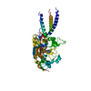
PDBj- Assembly
| Deposited unit | 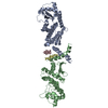
| ||||||||
|---|---|---|---|---|---|---|---|---|---|
| 1 | 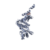
| ||||||||
| 2 | 
| ||||||||
| Unit cell |
|
-Components
| #1: Protein | Mass: 37832.027 Da / Num. of mol.: 2 Source method: isolated from a genetically manipulated source Source: (gene. exp.) Salmonella typhimurium (strain LT2 / SGSC1412 / ATCC 700720) (bacteria)Strain: LT2 / SGSC1412 / ATCC 700720 / Gene: sopD2, STM0972 / Plasmid: pGEX-4T1 / Production host: #2: Protein/peptide | Mass: 954.168 Da / Num. of mol.: 2 Source method: isolated from a genetically manipulated source Source: (gene. exp.) Salmonella enterica subsp. enterica serovar Typhimurium (bacteria)Production host: Has protein modification | Y | Sequence details | Author believes that chain C and chain D are part of the SopD2 protein. They are built based on the ...Author believes that chain C and chain D are part of the SopD2 protein. They are built based on the electron density maps but the side chains of these two segments are not clear due to the poor quality of the maps therefore residues identity can not be assigned unambiguously. Very likely these are part of the flexible N-terminal (residues 1-35) segment, i.e., chain C is part of the N-terminal of chain A and chain D is part of the N-terminal of chain B. | |
|---|
-Experimental details
-Experiment
| Experiment | Method: X-RAY DIFFRACTION / Number of used crystals: 1 |
|---|
- Sample preparation
| Crystal | Density Matthews: 2.76 Å3/Da / Density % sol: 55.44 % |
|---|---|
| Crystal grow | Temperature: 295 K / Method: microbatch / pH: 9.5 / Details: 0.1M CHES pH 9.5, 20% PEG 8000 |
-Data collection
| Diffraction | Mean temperature: 100 K |
|---|---|
| Diffraction source | Source: SYNCHROTRON / Site: CLSI / Beamline: 08ID-1 / Wavelength: 0.9795 Å |
| Detector | Type: MAR CCD 165 mm / Detector: CCD / Date: Aug 10, 2012 |
| Radiation | Protocol: SINGLE WAVELENGTH / Monochromatic (M) / Laue (L): M / Scattering type: x-ray |
| Radiation wavelength | Wavelength: 0.9795 Å / Relative weight: 1 |
| Reflection | Resolution: 3→50 Å / Num. obs: 16873 / % possible obs: 99.8 % / Redundancy: 3.8 % / Rmerge(I) obs: 0.059 / Net I/σ(I): 12.8 |
| Reflection shell | Resolution: 3→3.11 Å / Redundancy: 3.8 % / Rmerge(I) obs: 0.739 / % possible all: 100 |
- Processing
| Software |
| ||||||||||||||||||||||||||||||||||||||||||||||||||||||||||||||||||||||||||||||||||||||||||||||||||||
|---|---|---|---|---|---|---|---|---|---|---|---|---|---|---|---|---|---|---|---|---|---|---|---|---|---|---|---|---|---|---|---|---|---|---|---|---|---|---|---|---|---|---|---|---|---|---|---|---|---|---|---|---|---|---|---|---|---|---|---|---|---|---|---|---|---|---|---|---|---|---|---|---|---|---|---|---|---|---|---|---|---|---|---|---|---|---|---|---|---|---|---|---|---|---|---|---|---|---|---|---|---|
| Refinement | Method to determine structure: MOLECULAR REPLACEMENT Starting model: 5CPC Resolution: 3→50 Å / SU B: 62.231 / SU ML: 0.498 / Cross valid method: THROUGHOUT / σ(F): 0 / ESU R Free: 0.506 Details: U VALUES : WITH TLS ADDED HYDROGENS HAVE BEEN ADDED IN THE RIDING POSITIONS
| ||||||||||||||||||||||||||||||||||||||||||||||||||||||||||||||||||||||||||||||||||||||||||||||||||||
| Displacement parameters | Biso mean: 86.56 Å2
| ||||||||||||||||||||||||||||||||||||||||||||||||||||||||||||||||||||||||||||||||||||||||||||||||||||
| Refinement step | Cycle: LAST / Resolution: 3→50 Å
| ||||||||||||||||||||||||||||||||||||||||||||||||||||||||||||||||||||||||||||||||||||||||||||||||||||
| Refinement TLS params. | Method: refined / Refine-ID: X-RAY DIFFRACTION
| ||||||||||||||||||||||||||||||||||||||||||||||||||||||||||||||||||||||||||||||||||||||||||||||||||||
| Refinement TLS group |
|
