 Movie
Movie Controller


+ Open data

- Basic information
| Entry | Database: PDB / ID: 2lyn | ||||||
|---|---|---|---|---|---|---|---|
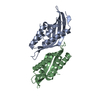
| Title | HIGH RESOLUTION STRUCTURE OF RED ABALONE LYSIN DIMER | ||||||
 Components Components | SPERM LYSIN | ||||||
 Keywords Keywords | CELL ADHESION / ABALONE LYSIN / FERTILIZATION PROTEIN / GAMETE RECOGNITION PROTEIN | ||||||
| Function / homology |  Function and homology information Function and homology information | ||||||
| Biological species |  Haliotis rufescens (red abalone) Haliotis rufescens (red abalone) | ||||||
| Method |  X-RAY DIFFRACTION / SYNCHROTRON / MOLECULAR REPLACEMENT / Resolution: 2.07 Å X-RAY DIFFRACTION / SYNCHROTRON / MOLECULAR REPLACEMENT / Resolution: 2.07 Å | ||||||
 Authors Authors | Kresge, N. / Vacquier, V.D. / Stout, C.D. | ||||||
 Citation Citation | Journal: Acta Crystallogr.,Sect.D / Year: 2000 Title: 1.35 and 2.07 A resolution structures of the red abalone sperm lysin monomer and dimer reveal features involved in receptor binding. Authors: Kresge, N. / Vacquier, V.D. / Stout, C.D. | ||||||
| History |
|
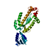
- Structure visualization
| Structure viewer | Molecule: MolmilJmol/JSmol |
|---|
- Downloads & links
-Download
| PDBx/mmCIF format | 2lyn.cif.gz | 122.4 KB | Display | PDBx/mmCIF format |
|---|---|---|---|---|
| PDB format | pdb2lyn.ent.gz | 97.2 KB | Display | PDB format |
| PDBx/mmJSON format | 2lyn.json.gz | Tree view | PDBx/mmJSON format | |
| Others |  Other downloads Other downloads |
-Validation report
| Arichive directory | https://data.pdbj.org/pub/pdb/validation_reports/ly/2lynftp://data.pdbj.org/pub/pdb/validation_reports/ly/2lyn | HTTPS FTP |
|---|
-Related structure data
| Related structure data |  2lisC 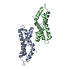 1lynS C: citing same article ( S: Starting model for refinement |
|---|---|
| Similar structure data |


-Links
 PDBj
PDBj- Assembly
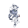
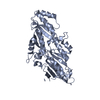
| Deposited unit | 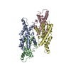
| |||||||||
|---|---|---|---|---|---|---|---|---|---|---|
| 1 | 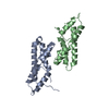
| |||||||||
| 2 | 
| |||||||||
| 3 |
| |||||||||
| Unit cell |
| |||||||||
| Components on special symmetry positions |
|
-Components
| #1: Protein | Mass: 16295.218 Da / Num. of mol.: 4 / Source method: isolated from a natural source / Source: (natural) Haliotis rufescens (red abalone) / Cell: SPERM / Organ: GONAD / Organelle: ACROSOME GRANULE / References: UniProt: P04552#2: Water | ChemComp-HOH / |  Mass: 18.015 Da / Num. of mol.: 316 / Source method: isolated from a natural source / Formula: H2O Mass: 18.015 Da / Num. of mol.: 316 / Source method: isolated from a natural source / Formula: H2O |
|---|
-Experimental details
-Experiment
| Experiment | Method: X-RAY DIFFRACTION / Number of used crystals: 1 |
|---|
- Sample preparation
| Crystal | Density Matthews: 2.69 Å3/Da / Density % sol: 53.88 % | ||||||||||||||||||||||||||||||||||||||||||||||||||||||
|---|---|---|---|---|---|---|---|---|---|---|---|---|---|---|---|---|---|---|---|---|---|---|---|---|---|---|---|---|---|---|---|---|---|---|---|---|---|---|---|---|---|---|---|---|---|---|---|---|---|---|---|---|---|---|---|
| Crystal grow | pH: 4.5 Details: 0.75 M (NH4)2SO4, 20 MM NH4SCN, 52.5 MM SODIUM-CITRATE-BORIC ACID BUFFER PH 4.5, 0.1 MM EDTA, 0.02% OTGP | ||||||||||||||||||||||||||||||||||||||||||||||||||||||
| Crystal grow | *PLUS Temperature: 295 K / pH: 5.5 / Method: vapor diffusion | ||||||||||||||||||||||||||||||||||||||||||||||||||||||
| Components of the solutions | *PLUS
|
-Data collection
| Diffraction | Mean temperature: 100 K |
|---|---|
| Diffraction source | Source: SYNCHROTRON / Site: SSRL  / Beamline: BL9-1 / Wavelength: 0.98 / Beamline: BL9-1 / Wavelength: 0.98 |
| Detector | Type: MARRESEARCH / Detector: IMAGE PLATE / Date: May 1, 1998 |
| Radiation | Protocol: SINGLE WAVELENGTH / Monochromatic (M) / Laue (L): M / Scattering type: x-ray |
| Radiation wavelength | Wavelength: 0.98 Å / Relative weight: 1 |
| Reflection | Resolution: 2.07→40.5 Å / Num. obs: 44137 / % possible obs: 99.1 % / Observed criterion σ(I): 2 / Redundancy: 3.6 % / Rsym value: 0.087 / Net I/σ(I): 4.8 |
| Reflection shell | Resolution: 2.07→2.18 Å / Redundancy: 2.5 % / Mean I/σ(I) obs: 2.8 / Rsym value: 0.256 / % possible all: 94.6 |
| Reflection | *PLUS Redundancy: 6.9 % / Num. measured all: 948262 / Rmerge(I) obs: 0.087 |
| Reflection shell | *PLUS % possible obs: 94.6 % / Redundancy: 4.5 % / Rmerge(I) obs: 0.256 |
- Processing
| Software |
| |||||||||||||||||||||||||||||||||
|---|---|---|---|---|---|---|---|---|---|---|---|---|---|---|---|---|---|---|---|---|---|---|---|---|---|---|---|---|---|---|---|---|---|---|
| Refinement | Method to determine structure: MOLECULAR REPLACEMENT Starting model: PDB ENTRY 1LYN Resolution: 2.07→100 Å / Num. parameters: 18473 / Num. restraintsaints: 17746 / σ(F): 0 / Stereochemistry target values: ENGH AND HUBER
| |||||||||||||||||||||||||||||||||
| Solvent computation | Solvent model: MOEWS & KRETSINGER, J.MOL.BIOL.91(1973)201-228 | |||||||||||||||||||||||||||||||||
| Refine analyze | Num. disordered residues: 2 / Occupancy sum hydrogen: 0 / Occupancy sum non hydrogen: 4604 | |||||||||||||||||||||||||||||||||
| Refinement step | Cycle: LAST / Resolution: 2.07→100 Å
| |||||||||||||||||||||||||||||||||
| Refine LS restraints |
| |||||||||||||||||||||||||||||||||
| Software | *PLUS Name: SHELXL-96 / Classification: refinement | |||||||||||||||||||||||||||||||||
| Refinement | *PLUS Lowest resolution: 40.5 Å / Rfactor Rfree: 0.285 | |||||||||||||||||||||||||||||||||
| Solvent computation | *PLUS | |||||||||||||||||||||||||||||||||
| Displacement parameters | *PLUS | |||||||||||||||||||||||||||||||||
| Refine LS restraints | *PLUS
|
