 Movie
Movie Controller


+ Open data

- Basic information
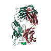
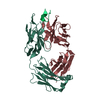
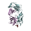
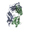
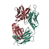
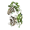
| Entry | Database: PDB / ID: 1q9k | ||||||
|---|---|---|---|---|---|---|---|
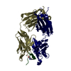
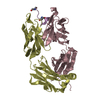
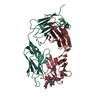
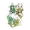
| Title | S25-2 Fab Unliganded 1 | ||||||
 Components Components |
| ||||||
 Keywords Keywords | IMMUNE SYSTEM / antigen-binding fragment / Fab / anti-carbohydrate / anti-LPS / antibody / immunoglobulin | ||||||
| Function / homology | Immunoglobulins / Immunoglobulin-like / Sandwich / Mainly Beta Function and homology information Function and homology information | ||||||
| Biological species |  | ||||||
| Method |  X-RAY DIFFRACTION / SYNCHROTRON / MOLECULAR REPLACEMENT / Resolution: 1.96 Å X-RAY DIFFRACTION / SYNCHROTRON / MOLECULAR REPLACEMENT / Resolution: 1.96 Å | ||||||
 Authors Authors | Nguyen, H.P. / Seto, N.O. / MacKenzie, C.R. / Brade, L. / Kosma, P. / Brade, H. / Evans, S.V. | ||||||
 Citation Citation | Journal: Nat.Struct.Biol. / Year: 2003 Title: Germline antibody recognition of distinct carbohydrate epitopes. Authors: Nguyen, H.P. / Seto, N.O. / MacKenzie, C.R. / Brade, L. / Kosma, P. / Brade, H. / Evans, S.V. | ||||||
| History |
| ||||||
| Remark 999 | SEQUENCE The sequence of the protein was not deposited into any sequence database. |
- Structure visualization
| Structure viewer | Molecule: MolmilJmol/JSmol |
|---|
- Downloads & links
-Download
| PDBx/mmCIF format | 1q9k.cif.gz | 106.1 KB | Display | PDBx/mmCIF format |
|---|---|---|---|---|
| PDB format | pdb1q9k.ent.gz | 80.1 KB | Display | PDB format |
| PDBx/mmJSON format | 1q9k.json.gz | Tree view | PDBx/mmJSON format | |
| Others |  Other downloads Other downloads |
-Validation report
| Arichive directory | https://data.pdbj.org/pub/pdb/validation_reports/q9/1q9kftp://data.pdbj.org/pub/pdb/validation_reports/q9/1q9k | HTTPS FTP |
|---|
-Related structure data
| Related structure data | 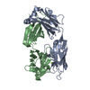 1q9lC 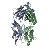 1q9oSC 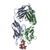 1q9wC  3sy0C  3t4yC  3t65C 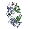 3t77C C: citing same article ( S: Starting model for refinement |
|---|---|
| Similar structure data |


-Links
 PDBj
PDBj

- Assembly
| Deposited unit | 
| ||||||||
|---|---|---|---|---|---|---|---|---|---|
| 1 |
| ||||||||
| Unit cell |
|
-Components
| #1: Antibody | Mass: 24242.898 Da / Num. of mol.: 1 / Fragment: Light Chain kappa / Source method: isolated from a natural source / Source: (natural) | ||||
|---|---|---|---|---|---|
| #2: Antibody | Mass: 24110.967 Da / Num. of mol.: 1 / Fragment: Heavy Chain g1 / Source method: isolated from a natural source / Source: (natural) | ||||
| #3: Chemical | ChemComp-MG /   Mass: 24.305 Da / Num. of mol.: 4 / Source method: obtained synthetically / Formula: Mg Mass: 24.305 Da / Num. of mol.: 4 / Source method: obtained synthetically / Formula: Mg#4: Water | ChemComp-HOH / |  Mass: 18.015 Da / Num. of mol.: 269 / Source method: isolated from a natural source / Formula: H2O Mass: 18.015 Da / Num. of mol.: 269 / Source method: isolated from a natural source / Formula: H2OHas protein modification | Y | |
-Experimental details
-Experiment
| Experiment | Method: X-RAY DIFFRACTION / Number of used crystals: 1 |
|---|
- Sample preparation
| Crystal | Density Matthews: 2.33 Å3/Da / Density % sol: 47.19 % | ||||||||||||||||||||||||||||||||||||||||||||||||||||||||||||||||||||||
|---|---|---|---|---|---|---|---|---|---|---|---|---|---|---|---|---|---|---|---|---|---|---|---|---|---|---|---|---|---|---|---|---|---|---|---|---|---|---|---|---|---|---|---|---|---|---|---|---|---|---|---|---|---|---|---|---|---|---|---|---|---|---|---|---|---|---|---|---|---|---|---|
| Crystal grow | Temperature: 293 K / Method: vapor diffusion, hanging drop / pH: 7 Details: zinc acetate, magnesium chloride, PEG 4000, PEG 8000, cacodylate, tris, pH 7, VAPOR DIFFUSION, HANGING DROP, temperature 293K | ||||||||||||||||||||||||||||||||||||||||||||||||||||||||||||||||||||||
| Crystal grow | *PLUS pH: 8 / Method: vapor diffusion, hanging drop | ||||||||||||||||||||||||||||||||||||||||||||||||||||||||||||||||||||||
| Components of the solutions | *PLUS
|
-Data collection
| Diffraction | Mean temperature: 100 K |
|---|---|
| Diffraction source | Source: SYNCHROTRON / Site: NSLS  / Beamline: X8C / Wavelength: 1.15 Å / Beamline: X8C / Wavelength: 1.15 Å |
| Detector | Type: ADSC QUANTUM 4 / Detector: CCD / Date: Mar 22, 2002 / Details: mirrors |
| Radiation | Protocol: SINGLE WAVELENGTH / Monochromatic (M) / Laue (L): M / Scattering type: x-ray |
| Radiation wavelength | Wavelength: 1.15 Å / Relative weight: 1 |
| Reflection | Resolution: 1.96→19.74 Å / Num. all: 31806 / Num. obs: 29982 / % possible obs: 90.1 % / Observed criterion σ(F): 0 / Observed criterion σ(I): 0 / Biso Wilson estimate: 23.1 Å2 / Limit h max: 48 / Limit h min: 0 / Limit k max: 56 / Limit k min: 0 / Limit l max: 20 / Limit l min: 0 / Observed criterion F max: 244216.73 / Observed criterion F min: 0.32 |
| Reflection shell | Resolution: 1.96→2.03 Å / % possible all: 87.5 |
| Reflection | *PLUS Lowest resolution: 20 Å / Num. obs: 31827 / % possible obs: 96.1 % / Num. measured all: 203789 / Rmerge(I) obs: 0.054 |
| Reflection shell | *PLUS % possible obs: 87.5 % / Num. unique obs: 2827 / Rmerge(I) obs: 0.328 / Mean I/σ(I) obs: 3.2 |
- Processing
| Software |
| ||||||||||||||||||||||||||||||||||||||||||||||||||||||||||||||||||||||||||||||||||||||||||||||||||||||||||||||
|---|---|---|---|---|---|---|---|---|---|---|---|---|---|---|---|---|---|---|---|---|---|---|---|---|---|---|---|---|---|---|---|---|---|---|---|---|---|---|---|---|---|---|---|---|---|---|---|---|---|---|---|---|---|---|---|---|---|---|---|---|---|---|---|---|---|---|---|---|---|---|---|---|---|---|---|---|---|---|---|---|---|---|---|---|---|---|---|---|---|---|---|---|---|---|---|---|---|---|---|---|---|---|---|---|---|---|---|---|---|---|---|
| Refinement | Method to determine structure: MOLECULAR REPLACEMENT Starting model: PDB entry 1Q9O, S45-18 Fab unliganded Resolution: 1.96→19.74 Å / Rfactor Rfree error: 0.005 / Occupancy max: 1 / Occupancy min: 1 / Cross valid method: THROUGHOUT / σ(F): 0 / Stereochemistry target values: Engh & Huber
| ||||||||||||||||||||||||||||||||||||||||||||||||||||||||||||||||||||||||||||||||||||||||||||||||||||||||||||||
| Solvent computation | Solvent model: CNS bulk solvent model used / Bsol: 67.8721 Å2 / ksol: 0.348868 e/Å3 | ||||||||||||||||||||||||||||||||||||||||||||||||||||||||||||||||||||||||||||||||||||||||||||||||||||||||||||||
| Displacement parameters | Biso max: 99.54 Å2 / Biso mean: 40.2 Å2 / Biso min: 18.64 Å2
| ||||||||||||||||||||||||||||||||||||||||||||||||||||||||||||||||||||||||||||||||||||||||||||||||||||||||||||||
| Refine analyze |
| ||||||||||||||||||||||||||||||||||||||||||||||||||||||||||||||||||||||||||||||||||||||||||||||||||||||||||||||
| Refinement step | Cycle: LAST / Resolution: 1.96→19.74 Å
| ||||||||||||||||||||||||||||||||||||||||||||||||||||||||||||||||||||||||||||||||||||||||||||||||||||||||||||||
| Refine LS restraints |
| ||||||||||||||||||||||||||||||||||||||||||||||||||||||||||||||||||||||||||||||||||||||||||||||||||||||||||||||
| LS refinement shell | Refine-ID: X-RAY DIFFRACTION / Total num. of bins used: 10
| ||||||||||||||||||||||||||||||||||||||||||||||||||||||||||||||||||||||||||||||||||||||||||||||||||||||||||||||
| Xplor file |
| ||||||||||||||||||||||||||||||||||||||||||||||||||||||||||||||||||||||||||||||||||||||||||||||||||||||||||||||
| Software | *PLUS Name: CNS / Classification: refinement | ||||||||||||||||||||||||||||||||||||||||||||||||||||||||||||||||||||||||||||||||||||||||||||||||||||||||||||||
| Refinement | *PLUS Lowest resolution: 20 Å / Rfactor Rfree: 0.275 / Rfactor Rwork: 0.223 | ||||||||||||||||||||||||||||||||||||||||||||||||||||||||||||||||||||||||||||||||||||||||||||||||||||||||||||||
| Solvent computation | *PLUS | ||||||||||||||||||||||||||||||||||||||||||||||||||||||||||||||||||||||||||||||||||||||||||||||||||||||||||||||
| Displacement parameters | *PLUS | ||||||||||||||||||||||||||||||||||||||||||||||||||||||||||||||||||||||||||||||||||||||||||||||||||||||||||||||
| Refine LS restraints | *PLUS Type: c_bond_d / Dev ideal: 0.01 |
