 ムービー
ムービー コントローラー


+ データを開く

- 基本情報
基本情報
| 登録情報 | データベース: PDB / ID: 1q9o | ||||||
|---|---|---|---|---|---|---|---|
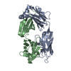
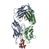
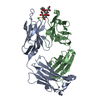
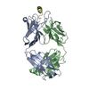
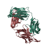
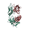
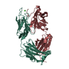
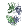
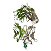
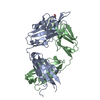
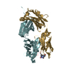
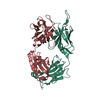
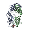
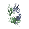
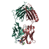
| タイトル | S45-18 Fab Unliganded | ||||||
 要素 要素 |
| ||||||
 キーワード キーワード | IMMUNE SYSTEM / antigen-binding fragment / Fab / anti-carbohydrate / anti-LPS / antibody / immunoglobulin | ||||||
| 機能・相同性 |  機能・相同性情報 機能・相同性情報alpha-beta T cell receptor complex / IgG immunoglobulin complex / immunoglobulin complex / B cell differentiation / adaptive immune response / extracellular region / metal ion binding / plasma membrane 類似検索 - 分子機能 | ||||||
| 生物種 |  | ||||||
| 手法 |  X線回折 / シンクロトロン / 分子置換 / 解像度: 1.79 Å X線回折 / シンクロトロン / 分子置換 / 解像度: 1.79 Å | ||||||
 データ登録者 データ登録者 | Nguyen, H.P. / Seto, N.O. / MacKenzie, C.R. / Brade, L. / Kosma, P. / Brade, H. / Evans, S.V. | ||||||
 引用 引用 | ジャーナル: Nat.Struct.Biol. / 年: 2003 タイトル: Germline antibody recognition of distinct carbohydrate epitopes. 著者: Nguyen, H.P. / Seto, N.O. / MacKenzie, C.R. / Brade, L. / Kosma, P. / Brade, H. / Evans, S.V. | ||||||
| 履歴 |
| ||||||
| Remark 999 | SEQUENCE The sequence of the protein was not deposited into any sequence database. |
- 構造の表示
構造の表示
| 構造ビューア | 分子: MolmilJmol/JSmol |
|---|
- ダウンロードとリンク
-ダウンロード
| PDBx/mmCIF形式 | 1q9o.cif.gz | 192.6 KB | 表示 | PDBx/mmCIF形式 |
|---|---|---|---|---|
| PDB形式 | pdb1q9o.ent.gz | 151.5 KB | 表示 | PDB形式 |
| PDBx/mmJSON形式 | 1q9o.json.gz | ツリー表示 | PDBx/mmJSON形式 | |
| その他 |  その他のダウンロード その他のダウンロード |
-検証レポート
| アーカイブディレクトリ | https://data.pdbj.org/pub/pdb/validation_reports/q9/1q9oftp://data.pdbj.org/pub/pdb/validation_reports/q9/1q9o | HTTPS FTP |
|---|
-関連構造データ


-リンク
 PDBj
PDBj


- 集合体
集合体
| 登録構造単位 | 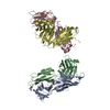
| ||||||||
|---|---|---|---|---|---|---|---|---|---|
| 1 | 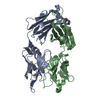
| ||||||||
| 2 | 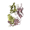
| ||||||||
| 単位格子 |
|
-要素
| #1: 抗体 | 分子量: 24445.398 Da / 分子数: 2 / 断片: Fab1 Heavy chain g1 / 由来タイプ: 天然 / 由来: (天然) #2: 抗体 | 分子量: 24306.039 Da / 分子数: 2 / 断片: Fab1 Light chain kappa / 由来タイプ: 天然 / 由来: (天然) #3: 化合物 | ChemComp-MG /   分子量: 24.305 Da / 分子数: 4 / 由来タイプ: 合成 / 式: Mg 分子量: 24.305 Da / 分子数: 4 / 由来タイプ: 合成 / 式: Mg#4: 水 | ChemComp-HOH / |  分子量: 18.015 Da / 分子数: 525 / 由来タイプ: 天然 / 式: H2O 分子量: 18.015 Da / 分子数: 525 / 由来タイプ: 天然 / 式: H2OHas protein modification | Y | |
|---|
-実験情報
-実験
| 実験 | 手法: X線回折 / 使用した結晶の数: 1 |
|---|
- 試料調製
試料調製
| 結晶 | マシュー密度: 2.34 Å3/Da / 溶媒含有率: 47.46 % | ||||||||||||||||||||||||||||||||||||||||||
|---|---|---|---|---|---|---|---|---|---|---|---|---|---|---|---|---|---|---|---|---|---|---|---|---|---|---|---|---|---|---|---|---|---|---|---|---|---|---|---|---|---|---|---|
| 結晶化 | 温度: 293 K / 手法: 蒸気拡散法, ハンギングドロップ法 / pH: 7.8 詳細: potassium phosphate, PEG 8000, pH 7.8, VAPOR DIFFUSION, HANGING DROP, temperature 293K | ||||||||||||||||||||||||||||||||||||||||||
| 結晶化 | *PLUS 温度: 295 K / 手法: 蒸気拡散法, ハンギングドロップ法 / 詳細: Nguyen, H.P., (2001) Acta Cryst., D57, 1872. | ||||||||||||||||||||||||||||||||||||||||||
| 溶液の組成 | *PLUS
|
-データ収集
| 回折 | 平均測定温度: 100 K |
|---|---|
| 放射光源 | 由来: シンクロトロン / サイト: NSLS  / ビームライン: X8C / 波長: 1.15 Å / ビームライン: X8C / 波長: 1.15 Å |
| 検出器 | タイプ: ADSC QUANTUM 4 / 検出器: CCD / 日付: 2002年2月5日 / 詳細: mirrors |
| 放射 | モノクロメーター: SI 111 CHANNEL / プロトコル: SINGLE WAVELENGTH / 単色(M)・ラウエ(L): M / 散乱光タイプ: x-ray |
| 放射波長 | 波長: 1.15 Å / 相対比: 1 |
| 反射 | 解像度: 1.79→19.18 Å / Num. all: 75932 / Num. obs: 74454 / % possible obs: 88.6 % / Observed criterion σ(F): 0 / Observed criterion σ(I): 0 / Biso Wilson estimate: 21.1 Å2 / Limit h max: 38 / Limit h min: -42 / Limit k max: 95 / Limit k min: -42 / Limit l max: 43 / Limit l min: 0 / Observed criterion F max: 240848.79 / Observed criterion F min: 0.32 |
| 反射 シェル | 解像度: 1.79→1.85 Å / % possible all: 78.5 |
| 反射 | *PLUS 最低解像度: 20 Å / Num. obs: 75932 / % possible obs: 90.4 % / Observed criterion σ(F): 0 / Num. measured all: 234850 / Rmerge(I) obs: 0.03 |
| 反射 シェル | *PLUS % possible obs: 78.5 % / Num. unique obs: 6553 / Rmerge(I) obs: 0.133 / Mean I/σ(I) obs: 6.9 |
- 解析
解析
| ソフトウェア |
| ||||||||||||||||||||||||||||||||||||||||||||||||||||||||||||||||||||||||||||||||||||||||||||||||||||||||||||||
|---|---|---|---|---|---|---|---|---|---|---|---|---|---|---|---|---|---|---|---|---|---|---|---|---|---|---|---|---|---|---|---|---|---|---|---|---|---|---|---|---|---|---|---|---|---|---|---|---|---|---|---|---|---|---|---|---|---|---|---|---|---|---|---|---|---|---|---|---|---|---|---|---|---|---|---|---|---|---|---|---|---|---|---|---|---|---|---|---|---|---|---|---|---|---|---|---|---|---|---|---|---|---|---|---|---|---|---|---|---|---|---|
| 精密化 | 構造決定の手法: 分子置換 開始モデル: Fv domain of YST 9.1 Fab unliganded (PDB entry 1MAM) 解像度: 1.79→19.18 Å / Rfactor Rfree error: 0.003 / Occupancy max: 1 / Occupancy min: 1 / 交差検証法: THROUGHOUT / σ(F): 0 / 立体化学のターゲット値: Engh & Huber
| ||||||||||||||||||||||||||||||||||||||||||||||||||||||||||||||||||||||||||||||||||||||||||||||||||||||||||||||
| 溶媒の処理 | 溶媒モデル: CNS bulk solvent model used / Bsol: 43.4159 Å2 / ksol: 0.327425 e/Å3 | ||||||||||||||||||||||||||||||||||||||||||||||||||||||||||||||||||||||||||||||||||||||||||||||||||||||||||||||
| 原子変位パラメータ | Biso max: 84.92 Å2 / Biso mean: 30.11 Å2 / Biso min: 14.35 Å2
| ||||||||||||||||||||||||||||||||||||||||||||||||||||||||||||||||||||||||||||||||||||||||||||||||||||||||||||||
| Refine analyze |
| ||||||||||||||||||||||||||||||||||||||||||||||||||||||||||||||||||||||||||||||||||||||||||||||||||||||||||||||
| 精密化ステップ | サイクル: LAST / 解像度: 1.79→19.18 Å
| ||||||||||||||||||||||||||||||||||||||||||||||||||||||||||||||||||||||||||||||||||||||||||||||||||||||||||||||
| 拘束条件 |
| ||||||||||||||||||||||||||||||||||||||||||||||||||||||||||||||||||||||||||||||||||||||||||||||||||||||||||||||
| LS精密化 シェル | Refine-ID: X-RAY DIFFRACTION / Total num. of bins used: 10
| ||||||||||||||||||||||||||||||||||||||||||||||||||||||||||||||||||||||||||||||||||||||||||||||||||||||||||||||
| Xplor file |
| ||||||||||||||||||||||||||||||||||||||||||||||||||||||||||||||||||||||||||||||||||||||||||||||||||||||||||||||
| ソフトウェア | *PLUS 名称: CNS / 分類: refinement | ||||||||||||||||||||||||||||||||||||||||||||||||||||||||||||||||||||||||||||||||||||||||||||||||||||||||||||||
| 精密化 | *PLUS 最低解像度: 20 Å / Rfactor Rwork: 0.22 | ||||||||||||||||||||||||||||||||||||||||||||||||||||||||||||||||||||||||||||||||||||||||||||||||||||||||||||||
| 溶媒の処理 | *PLUS | ||||||||||||||||||||||||||||||||||||||||||||||||||||||||||||||||||||||||||||||||||||||||||||||||||||||||||||||
| 原子変位パラメータ | *PLUS | ||||||||||||||||||||||||||||||||||||||||||||||||||||||||||||||||||||||||||||||||||||||||||||||||||||||||||||||
| 拘束条件 | *PLUS タイプ: c_bond_d / Dev ideal: 0.007 |
