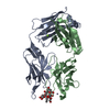
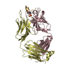
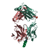
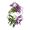
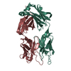
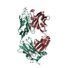
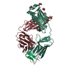
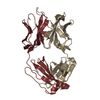
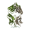
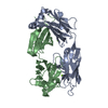
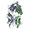
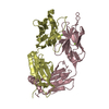
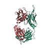
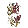
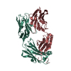
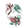
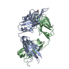
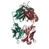
[English] 日本語
Yorodumi
- PDB-5mo3: Crystal structure of DC8E8 Fab in the complex with a 14-mer tau p... -

+
Open data


ID or keywords:

Loading...

-
Basic information

Entry
Database: PDB / ID: 5mo3
TitleCrystal structure of DC8E8 Fab in the complex with a 14-mer tau peptide at pH 8.5
Components
  • (Fab of monoclonal antibody, ...) x 2
  • Microtubule-associated protein tau
KeywordsIMMUNE SYSTEM / Fab / complex
Function / homology
Function and homology information


plus-end-directed organelle transport along microtubule / histone-dependent DNA binding / negative regulation of establishment of protein localization to mitochondrion / neurofibrillary tangle / microtubule lateral binding / axonal transport / positive regulation of protein localization to synapse / main axon / phosphatidylinositol bisphosphate binding / regulation of long-term synaptic depression ...plus-end-directed organelle transport along microtubule / histone-dependent DNA binding / negative regulation of establishment of protein localization to mitochondrion / neurofibrillary tangle / microtubule lateral binding / axonal transport / positive regulation of protein localization to synapse / main axon / phosphatidylinositol bisphosphate binding / regulation of long-term synaptic depression / tubulin complex / negative regulation of tubulin deacetylation / generation of neurons / regulation of chromosome organization / rRNA metabolic process / axonal transport of mitochondrion / regulation of mitochondrial fission / axon development / central nervous system neuron development / intracellular distribution of mitochondria / regulation of microtubule polymerization / microtubule polymerization / lipoprotein particle binding / minor groove of adenine-thymine-rich DNA binding / dynactin binding / negative regulation of mitochondrial membrane potential / apolipoprotein binding / glial cell projection / axolemma / protein polymerization / negative regulation of mitochondrial fission / Caspase-mediated cleavage of cytoskeletal proteins / regulation of microtubule polymerization or depolymerization / positive regulation of axon extension / neurofibrillary tangle assembly / Activation of AMPK downstream of NMDARs / synapse assembly / regulation of cellular response to heat / supramolecular fiber organization / positive regulation of protein localization / regulation of calcium-mediated signaling / somatodendritic compartment / cellular response to brain-derived neurotrophic factor stimulus / cytoplasmic microtubule organization / axon cytoplasm / positive regulation of microtubule polymerization / stress granule assembly / phosphatidylinositol binding / regulation of microtubule cytoskeleton organization / nuclear periphery / protein phosphatase 2A binding / positive regulation of superoxide anion generation / cellular response to reactive oxygen species / astrocyte activation / Hsp90 protein binding / microglial cell activation / cellular response to nerve growth factor stimulus / response to lead ion / synapse organization / PKR-mediated signaling / protein homooligomerization / regulation of synaptic plasticity / SH3 domain binding / memory / microtubule cytoskeleton organization / cytoplasmic ribonucleoprotein granule / neuron projection development / cell-cell signaling / single-stranded DNA binding / protein-folding chaperone binding / actin binding / cellular response to heat / microtubule cytoskeleton / cell body / growth cone / double-stranded DNA binding / microtubule binding / protein-macromolecule adaptor activity / dendritic spine / sequence-specific DNA binding / microtubule / amyloid fibril formation / learning or memory / neuron projection / regulation of autophagy / membrane raft / axon / negative regulation of gene expression / neuronal cell body / dendrite / DNA damage response / protein kinase binding / enzyme binding / mitochondrion / DNA binding / RNA binding / extracellular region / identical protein binding / nucleus / plasma membrane
Similarity search - Function
Microtubule-associated protein Tau / Microtubule associated protein, tubulin-binding repeat / Tau and MAP protein, tubulin-binding repeat / Tau and MAP proteins tubulin-binding repeat signature. / Tau and MAP proteins tubulin-binding repeat profile. / : / Immunoglobulins / Immunoglobulin-like / Sandwich / Mainly Beta
Similarity search - Domain/homology
trehalose / TRIETHYLENE GLYCOL / Microtubule-associated protein tau
Similarity search - Component
Biological speciesMus musculus (house mouse)
Homo sapiens (human)
MethodX-RAY DIFFRACTION / SYNCHROTRON / MOLECULAR REPLACEMENT / molecular replacement / Resolution: 1.69 Å
AuthorsSkrabana, R. / Novak, M.
CitationJournal: To be published
Title: Crystal structure of DC8E8 Fab in the complex with a 14-mer tau peptide at pH 8.5
Authors: Skrabana, R. / Novak, M.
History
DepositionDec 13, 2016Deposition site: PDBE / Processing site: PDBE
Revision 1.0May 2, 2018Provider: repository / Type: Initial release
Revision 2.0Mar 11, 2020Group: Polymer sequence / Category: entity_poly / Item: _entity_poly.pdbx_seq_one_letter_code_can
Revision 3.0Jul 29, 2020Group: Atomic model / Data collection ...Atomic model / Data collection / Derived calculations / Non-polymer description / Structure summary
Category: atom_site / chem_comp ...atom_site / chem_comp / entity / entity_name_com / pdbx_branch_scheme / pdbx_chem_comp_identifier / pdbx_entity_branch / pdbx_entity_branch_descriptor / pdbx_entity_branch_link / pdbx_entity_branch_list / pdbx_entity_nonpoly / pdbx_molecule_features / pdbx_nonpoly_scheme / struct_asym / struct_conn / struct_site / struct_site_gen
Item: _atom_site.B_iso_or_equiv / _atom_site.Cartn_x ..._atom_site.B_iso_or_equiv / _atom_site.Cartn_x / _atom_site.Cartn_y / _atom_site.Cartn_z / _atom_site.auth_asym_id / _atom_site.auth_atom_id / _atom_site.auth_comp_id / _atom_site.auth_seq_id / _atom_site.label_asym_id / _atom_site.label_atom_id / _atom_site.label_comp_id / _atom_site.label_entity_id / _atom_site.type_symbol / _chem_comp.formula / _chem_comp.formula_weight / _chem_comp.id / _chem_comp.mon_nstd_flag / _chem_comp.name / _chem_comp.pdbx_synonyms / _chem_comp.type / _entity.formula_weight / _entity.pdbx_description / _entity.pdbx_number_of_molecules / _entity.src_method / _entity.type / _struct_asym.entity_id
Description: Carbohydrate remediation / Provider: repository / Type: Remediation
Revision 3.1Jan 17, 2024Group: Data collection / Database references ...Data collection / Database references / Refinement description / Structure summary
Category: chem_comp / chem_comp_atom ...chem_comp / chem_comp_atom / chem_comp_bond / database_2 / pdbx_initial_refinement_model
Item: _chem_comp.pdbx_synonyms / _database_2.pdbx_DOI / _database_2.pdbx_database_accession
Revision 3.2Nov 6, 2024Group: Structure summary / Category: pdbx_entry_details / pdbx_modification_feature

-
Structure visualization

Structure viewerMolecule:
MolmilJmol/JSmol

Downloads & links

-
Assembly

Deposited unit
A: Microtubule-associated protein tau
H: Fab of monoclonal antibody, heavy chain
L: Fab of monoclonal antibody, light chain
hetero molecules


Theoretical massNumber of molelcules
Total (without water)50,3888
Polymers49,4453
Non-polymers9435
Water7,260403
1


  • Idetical with deposited unit
  • defined by author&software
TypeNameSymmetry operationNumber
identity operation1_555x,y,z1
Buried area6260 Å2
ΔGint-17 kcal/mol
Surface area20070 Å2
MethodPISA
Unit cell
Length a, b, c (Å)58.728, 60.128, 69.315
Angle α, β, γ (deg.)90.000, 109.250, 90.000
Int Tables number4
Space group name H-MP1211

-
Components

-
Antibody , 2 types, 2 molecules HL

#2: Antibody Fab of monoclonal antibody, heavy chain


Mass: 23800.617 Da / Num. of mol.: 1 / Source method: isolated from a natural source / Source: (natural) Mus musculus (house mouse)
#3: Antibody Fab of monoclonal antibody, light chain


Mass: 24172.787 Da / Num. of mol.: 1 / Source method: isolated from a natural source / Source: (natural) Mus musculus (house mouse)

-
Protein/peptide / Sugars , 2 types, 2 molecules A

#1: Protein/peptide Microtubule-associated protein tau / Neurofibrillary tangle protein / Paired helical filament-tau / PHF-tau


Mass: 1471.722 Da / Num. of mol.: 1 / Fragment: UNP residues 615-628 / Source method: obtained synthetically / Source: (synth.) Homo sapiens (human) / References: UniProt: P10636
#4: Polysaccharide alpha-D-glucopyranose-(1-1)-alpha-D-glucopyranose / trehalose


Type: oligosaccharide, Oligosaccharide / Class: Nutrient / Mass: 342.297 Da / Num. of mol.: 1
Source method: isolated from a genetically manipulated source
Details: oligosaccharide with reducing-end-to-reducing-end glycosidic bond
References: trehalose
DescriptorTypeProgram
DGlcpa1-1DGlcpaGlycam Condensed SequenceGMML 1.0
WURCS=2.0/1,2,1/[a2122h-1a_1-5]/1-1/a1-b1WURCSPDB2Glycan 1.1.0
[][a-D-Glcp]{[(1+1)][a-D-Glcp]{}}LINUCSPDB-CARE

-
Non-polymers , 2 types, 407 molecules

#5: Chemical
ChemComp-PGE / TRIETHYLENE GLYCOL


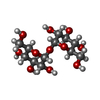
Mass: 150.173 Da / Num. of mol.: 4 / Source method: isolated from a natural source / Formula: C6H14O4
#6: Water ChemComp-HOH / water


Mass: 18.015 Da / Num. of mol.: 403 / Source method: isolated from a natural source / Formula: H2O

-
Details

Has protein modificationY

-
Experimental details

-
Experiment

ExperimentMethod: X-RAY DIFFRACTION / Number of used crystals: 1

-
Sample preparation

CrystalDensity Matthews: 2.37 Å3/Da / Density % sol: 48.14 %
Crystal growTemperature: 295 K / Method: vapor diffusion, hanging drop / pH: 8.5 / Details: 30% PEG 4000, 0.1 M Tris, 0.2 M LiSO4

-
Data collection

DiffractionMean temperature: 100 K
Diffraction sourceSource: SYNCHROTRON / Site: SLS / Beamline: X06DA / Wavelength: 0.9999 Å
DetectorType: MARMOSAIC 225 mm CCD / Detector: CCD / Date: Dec 5, 2009
RadiationProtocol: SINGLE WAVELENGTH / Monochromatic (M) / Laue (L): M / Scattering type: x-ray
Radiation wavelengthWavelength: 0.9999 Å / Relative weight: 1
ReflectionResolution: 1.69→65.44 Å / Num. obs: 41553 / % possible obs: 81.1 % / Observed criterion σ(I): -3 / Redundancy: 3.12 % / Biso Wilson estimate: 34.473 Å2 / CC1/2: 0.999 / Rmerge(I) obs: 0.059 / Rrim(I) all: 0.071 / Χ2: 0.937 / Net I/σ(I): 13.83 / Num. measured all: 129645 / Scaling rejects: 1009
Reflection shell
Resolution (Å)Redundancy (%)Rmerge(I) obsMean I/σ(I) obsCC1/2Diffraction-ID% possible all
1.69-1.733.0951.3790.890.448151.3
1.73-1.783.1081.1211.170.528163
1.78-1.833.1050.9891.370.636179.7
1.83-1.893.1571.5161.010.696193.2
1.89-1.953.1521.1481.280.83714.9
1.95-2.023.1550.4343.170.91196
2.02-2.13.1490.2754.750.943196.4
2.1-2.183.1650.2275.710.959196.6
2.18-2.283.1080.3413.990.963151.6
2.28-2.393.1640.1617.640.98195.3
2.39-2.523.1330.10910.790.988197.4
2.52-2.673.150.07714.550.993197.3
2.67-2.863.1360.05718.870.996197.6
2.86-3.093.1390.0425.160.998198
3.09-3.383.0990.03131.40.998197.5
3.38-3.783.090.02835.930.998197.9
3.78-4.363.0520.02342.310.999198.3
4.36-5.343.0480.0247.310.999197.8
5.34-7.562.9780.02244.460.998198.1
7.56-65.442.8270.02150.10.998195

-
Phasing

PhasingMethod: molecular replacement

-
Processing

Software
NameVersionClassification
XSCALEdata scaling
REFMAC5.8.0151refinement
PDB_EXTRACT3.22data extraction
XDSdata reduction
PHASERphasing
RefinementMethod to determine structure: MOLECULAR REPLACEMENT
Starting model: 4OZ4

4oz4
PDB Unreleased entry


Resolution: 1.69→65.44 Å / Cor.coef. Fo:Fc: 0.967 / Cor.coef. Fo:Fc free: 0.95 / SU B: 4.042 / SU ML: 0.119 / Cross valid method: THROUGHOUT / σ(F): 0 / ESU R: 0.142 / ESU R Free: 0.136
Details: HYDROGENS HAVE BEEN ADDED IN THE RIDING POSITIONS U VALUES : REFINED INDIVIDUALLY
RfactorNum. reflection% reflectionSelection details
Rfree0.2347 2092 5 %RANDOM
Rwork0.1894 ---
obs0.1917 39462 81.15 %-
Solvent computationIon probe radii: 0.8 Å / Shrinkage radii: 0.8 Å / VDW probe radii: 1.2 Å
Displacement parametersBiso max: 103.33 Å2 / Biso mean: 29.157 Å2 / Biso min: 15.14 Å2
Baniso -1Baniso -2Baniso -3
1--0.74 Å2-0 Å2-1.84 Å2
2--2.65 Å2-0 Å2
3----0.51 Å2
Refinement stepCycle: final / Resolution: 1.69→65.44 Å
ProteinNucleic acidLigandSolventTotal
Num. atoms3404 0 63 403 3870
Biso mean--63.42 36.24 -
Num. residues----446
Refine LS restraints
Refine-IDTypeDev idealDev ideal targetNumber
X-RAY DIFFRACTIONr_bond_refined_d0.0110.023608
X-RAY DIFFRACTIONr_bond_other_d0.0020.023295
X-RAY DIFFRACTIONr_angle_refined_deg1.581.964899
X-RAY DIFFRACTIONr_angle_other_deg0.9813.0027657
X-RAY DIFFRACTIONr_dihedral_angle_1_deg7.0465.022451
X-RAY DIFFRACTIONr_dihedral_angle_2_deg37.83124.234137
X-RAY DIFFRACTIONr_dihedral_angle_3_deg13.94115576
X-RAY DIFFRACTIONr_dihedral_angle_4_deg16.2131514
X-RAY DIFFRACTIONr_chiral_restr0.0930.2554
X-RAY DIFFRACTIONr_gen_planes_refined0.0060.0213982
X-RAY DIFFRACTIONr_gen_planes_other0.0010.02786
LS refinement shellResolution: 1.69→1.734 Å / Rfactor Rfree error: 0 / Total num. of bins used: 20
RfactorNum. reflection% reflection
Rfree0.398 83 -
Rwork0.337 1851 -
all-1934 -
obs--51.11 %

+
About Yorodumi

-
News

-
Feb 9, 2022. New format data for meta-information of EMDB entries

New format data for meta-information of EMDB entries

  • Version 3 of the EMDB header file is now the official format.
  • The previous official version 1.9 will be removed from the archive.

Related info.:EMDB header

External links:wwPDB to switch to version 3 of the EMDB data model

-
Aug 12, 2020. Covid-19 info

Covid-19 info

URL: https://pdbj.org/emnavi/covid19.php

New page: Covid-19 featured information page in EM Navigator.

Related info.:Covid-19 info / Mar 5, 2020. Novel coronavirus structure data

+
Mar 5, 2020. Novel coronavirus structure data

Novel coronavirus structure data

Related info.:Yorodumi Speices / Aug 12, 2020. Covid-19 info

External links:COVID-19 featured content - PDBj / Molecule of the Month (242):Coronavirus Proteases

+
Jan 31, 2019. EMDB accession codes are about to change! (news from PDBe EMDB page)

EMDB accession codes are about to change! (news from PDBe EMDB page)

  • The allocation of 4 digits for EMDB accession codes will soon come to an end. Whilst these codes will remain in use, new EMDB accession codes will include an additional digit and will expand incrementally as the available range of codes is exhausted. The current 4-digit format prefixed with “EMD-” (i.e. EMD-XXXX) will advance to a 5-digit format (i.e. EMD-XXXXX), and so on. It is currently estimated that the 4-digit codes will be depleted around Spring 2019, at which point the 5-digit format will come into force.
  • The EM Navigator/Yorodumi systems omit the EMD- prefix.

Related info.:Q: What is EMD? / ID/Accession-code notation in Yorodumi/EM Navigator

External links:EMDB Accession Codes are Changing Soon! / Contact to PDBj

+
Jul 12, 2017. Major update of PDB

Major update of PDB

  • wwPDB released updated PDB data conforming to the new PDBx/mmCIF dictionary.
  • This is a major update changing the version number from 4 to 5, and with Remediation, in which all the entries are updated.
  • In this update, many items about electron microscopy experimental information are reorganized (e.g. em_software).
  • Now, EM Navigator and Yorodumi are based on the updated data.

External links:wwPDB Remediation / Enriched Model Files Conforming to OneDep Data Standards Now Available in the PDB FTP Archive

-
Yorodumi

Thousand views of thousand structures

  • Yorodumi is a browser for structure data from EMDB, PDB, SASBDB, etc.
  • This page is also the successor to EM Navigator detail page, and also detail information page/front-end page for Omokage search.
  • The word "yorodu" (or yorozu) is an old Japanese word meaning "ten thousand". "mi" (miru) is to see.

Related info.:EMDB / PDB / SASBDB / Comparison of 3 databanks / Yorodumi Search / Aug 31, 2016. New EM Navigator & Yorodumi / Yorodumi Papers / Jmol/JSmol / Function and homology information / Changes in new EM Navigator and Yorodumi

Read more