 Movie
Movie Controller

[English] 日本語
 Yorodumi
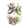
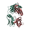
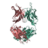
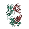
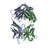
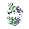
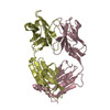
- PDB-1psk: THE CRYSTAL STRUCTURE OF AN FAB FRAGMENT THAT BINDS TO THE MELANO... -
+ Open data

- Basic information
| Entry | Database: PDB / ID: 1psk | ||||||
|---|---|---|---|---|---|---|---|
| Title | THE CRYSTAL STRUCTURE OF AN FAB FRAGMENT THAT BINDS TO THE MELANOMA-ASSOCIATED GD2 GANGLIOSIDE | ||||||
 Components Components | (ANTIBODY) x 2 | ||||||
 Keywords Keywords | IMMUNOGLOBULIN / FAB / GD2-GANGLIOSIDE / CARBOHYDRATE / MELANOMA | ||||||
| Function / homology |  Function and homology information Function and homology informationalpha-beta T cell receptor complex / IgG immunoglobulin complex / B cell differentiation / adaptive immune response / extracellular region / plasma membrane Similarity search - Function | ||||||
| Biological species |  | ||||||
| Method |  X-RAY DIFFRACTION / MOLECULAR REPLACEMENT / Resolution: 2.8 Å X-RAY DIFFRACTION / MOLECULAR REPLACEMENT / Resolution: 2.8 Å | ||||||
 Authors Authors | Pichla, S.L. / Murali, R. / Burnett, R.M. | ||||||
 Citation Citation | Journal: J.Struct.Biol. / Year: 1997 Title: The crystal structure of a Fab fragment to the melanoma-associated GD2 ganglioside. Authors: Pichla, S.L. / Murali, R. / Burnett, R.M. #1: Journal: Acta Crystallogr.,Sect.D / Year: 1995Title: Preliminary Crystallographic Data for an Fab to the Melanoma-Associated Gd2 Ganglioside, and the Purification of a Soluble Form of This Antigen Authors: Pichla, S.L. / Murali, R. / Burnett, R.M. | ||||||
| History |
|
- Structure visualization
| Structure viewer | Molecule: MolmilJmol/JSmol |
|---|
- Downloads & links
-Download
| PDBx/mmCIF format | 1psk.cif.gz | 106.3 KB | Display | PDBx/mmCIF format |
|---|---|---|---|---|
| PDB format | pdb1psk.ent.gz | 81.8 KB | Display | PDB format |
| PDBx/mmJSON format | 1psk.json.gz | Tree view | PDBx/mmJSON format | |
| Others |  Other downloads Other downloads |
-Validation report
| Arichive directory | https://data.pdbj.org/pub/pdb/validation_reports/ps/1pskftp://data.pdbj.org/pub/pdb/validation_reports/ps/1psk | HTTPS FTP |
|---|
-Related structure data
| Related structure data |  2fbjS S: Starting model for refinement |
|---|---|
| Similar structure data |


-Links
 PDBj
PDBj


- Assembly
| Deposited unit | 
| ||||||||
|---|---|---|---|---|---|---|---|---|---|
| 1 |
| ||||||||
| Unit cell |
|
-Components
| #1: Antibody | Mass: 23209.670 Da / Num. of mol.: 1 / Fragment: FAB / Source method: isolated from a natural source / Source: (natural) |
|---|---|
| #2: Antibody | Mass: 22368.936 Da / Num. of mol.: 1 / Fragment: FAB / Source method: isolated from a natural source / Source: (natural) |
| #3: Water | ChemComp-HOH /  Mass: 18.015 Da / Num. of mol.: 34 / Source method: isolated from a natural source / Formula: H2O Mass: 18.015 Da / Num. of mol.: 34 / Source method: isolated from a natural source / Formula: H2O |
| Has protein modification | Y |
-Experimental details
-Experiment
| Experiment | Method: X-RAY DIFFRACTION / Number of used crystals: 6 |
|---|
- Sample preparation
| Crystal | Density Matthews: 2.49 Å3/Da / Density % sol: 51 % | ||||||||||||||||||||
|---|---|---|---|---|---|---|---|---|---|---|---|---|---|---|---|---|---|---|---|---|---|
| Crystal grow | pH: 7.5 / Details: 30% (W/V) PEG 4000, 1% MPD, TRIS BUFFER, PH 7.5 | ||||||||||||||||||||
| Crystal grow | *PLUS Method: unknown | ||||||||||||||||||||
| Components of the solutions | *PLUS
|
-Data collection
| Diffraction | Mean temperature: 287 K |
|---|---|
| Diffraction source | Source: ROTATING ANODE / Type: RIGAKU RUH2R / Wavelength: 1.5418 |
| Detector | Type: SIEMENS / Detector: AREA DETECTOR / Date: Oct 1, 1990 / Details: MIRRORS |
| Radiation | Monochromatic (M) / Laue (L): M / Scattering type: x-ray |
| Radiation wavelength | Wavelength: 1.5418 Å / Relative weight: 1 |
| Reflection | Resolution: 2.8→20 Å / Num. obs: 14677 / % possible obs: 96.1 % / Observed criterion σ(I): 0 / Redundancy: 2.3 % / Biso Wilson estimate: 19 Å2 / Rmerge(I) obs: 0.104 / Net I/σ(I): 2 |
| Reflection shell | Resolution: 2.8→3 Å / Redundancy: 2 % / Rmerge(I) obs: 0.147 / % possible all: 95.4 |
| Reflection | *PLUS Num. measured all: 33212 |
| Reflection shell | *PLUS % possible obs: 95.4 % / Num. unique obs: 2021 / Num. measured obs: 4012 |
- Processing
| Software |
| ||||||||||||||||||||||||||||||||||||||||||||||||||||||||||||
|---|---|---|---|---|---|---|---|---|---|---|---|---|---|---|---|---|---|---|---|---|---|---|---|---|---|---|---|---|---|---|---|---|---|---|---|---|---|---|---|---|---|---|---|---|---|---|---|---|---|---|---|---|---|---|---|---|---|---|---|---|---|
| Refinement | Method to determine structure: MOLECULAR REPLACEMENT Starting model: PDB ENTRY 2FBJ Resolution: 2.8→8 Å / Data cutoff high absF: 10000 / Data cutoff low absF: 0.001 / Isotropic thermal model: RESTRAINED / Cross valid method: THROUGHOUT / σ(F): 2
| ||||||||||||||||||||||||||||||||||||||||||||||||||||||||||||
| Displacement parameters | Biso mean: 20 Å2 | ||||||||||||||||||||||||||||||||||||||||||||||||||||||||||||
| Refine analyze | Luzzati coordinate error obs: 0.35 Å / Luzzati d res low obs: 8 Å | ||||||||||||||||||||||||||||||||||||||||||||||||||||||||||||
| Refinement step | Cycle: LAST / Resolution: 2.8→8 Å
| ||||||||||||||||||||||||||||||||||||||||||||||||||||||||||||
| Refine LS restraints |
| ||||||||||||||||||||||||||||||||||||||||||||||||||||||||||||
| LS refinement shell | Resolution: 2.8→2.93 Å / Total num. of bins used: 8
| ||||||||||||||||||||||||||||||||||||||||||||||||||||||||||||
| Xplor file |
| ||||||||||||||||||||||||||||||||||||||||||||||||||||||||||||
| Software | *PLUS Name: X-PLOR / Version: 3 / Classification: refinement | ||||||||||||||||||||||||||||||||||||||||||||||||||||||||||||
| Refinement | *PLUS | ||||||||||||||||||||||||||||||||||||||||||||||||||||||||||||
| Solvent computation | *PLUS | ||||||||||||||||||||||||||||||||||||||||||||||||||||||||||||
| Displacement parameters | *PLUS | ||||||||||||||||||||||||||||||||||||||||||||||||||||||||||||
| Refine LS restraints | *PLUS
| ||||||||||||||||||||||||||||||||||||||||||||||||||||||||||||
| LS refinement shell | *PLUS Rfactor obs: 0.29 |
