 Movie
Movie Controller

[English] 日本語
 Yorodumi
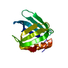
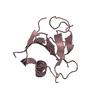
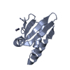
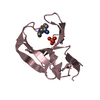
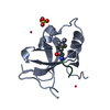
- PDB-1otd: STRONG HYDROGEN BONDS IN PHOTOACTIVE YELLOW PROTEIN AND THEIR ROL... -
+ Open data

- Basic information
| Entry | Database: PDB / ID: 1otd | ||||||
|---|---|---|---|---|---|---|---|
| Title | STRONG HYDROGEN BONDS IN PHOTOACTIVE YELLOW PROTEIN AND THEIR ROLE IN ITS PHOTOCYCLE | ||||||
 Components Components | Photoactive yellow protein | ||||||
 Keywords Keywords | SIGNALING PROTEIN / PYP | ||||||
| Function / homology |  Function and homology information Function and homology informationphotoreceptor activity / phototransduction / regulation of DNA-templated transcription / identical protein binding Similarity search - Function | ||||||
| Biological species |  Halorhodospira halophila (bacteria) Halorhodospira halophila (bacteria) | ||||||
| Method |  X-RAY DIFFRACTION / SYNCHROTRON / MOLECULAR REPLACEMENT / Resolution: 1.25 Å X-RAY DIFFRACTION / SYNCHROTRON / MOLECULAR REPLACEMENT / Resolution: 1.25 Å | ||||||
 Authors Authors | Anderson, S. / Crosson, S. / Moffat, K. | ||||||
 Citation Citation | Journal: Acta Crystallogr.,Sect.D / Year: 2004 Title: Short hydrogen bonds in photoactive yellow protein. Authors: Anderson, S. / Crosson, S. / Moffat, K. | ||||||
| History |
| ||||||
| Remark 999 | SEQRES Author states the sequence from the PYP was cloned from the strain SL-1 of ...SEQRES Author states the sequence from the PYP was cloned from the strain SL-1 of Ectothiorhodospira halophila, and there was no appropriate sequence database reference available at the time of processing. |
- Structure visualization
| Structure viewer | Molecule: MolmilJmol/JSmol |
|---|
- Downloads & links
-Download
| PDBx/mmCIF format | 1otd.cif.gz | 109.2 KB | Display | PDBx/mmCIF format |
|---|---|---|---|---|
| PDB format | pdb1otd.ent.gz | 82.3 KB | Display | PDB format |
| PDBx/mmJSON format | 1otd.json.gz | Tree view | PDBx/mmJSON format | |
| Others |  Other downloads Other downloads |
-Validation report
| Arichive directory | https://data.pdbj.org/pub/pdb/validation_reports/ot/1otdftp://data.pdbj.org/pub/pdb/validation_reports/ot/1otd | HTTPS FTP |
|---|
-Related structure data
| Related structure data |  1ot6C  1ot9C  1otaC 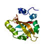 1otbC  1oteC  1otiC 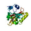 2phyS S: Starting model for refinement C: citing same article ( |
|---|---|
| Similar structure data |


-Links
 PDBj
PDBj


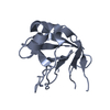
- Assembly
| Deposited unit | 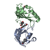
| ||||||||
|---|---|---|---|---|---|---|---|---|---|
| 1 | 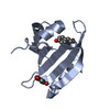
| ||||||||
| 2 | 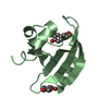
| ||||||||
| Unit cell |
|
-Components
| #1: Protein | Mass: 13886.557 Da / Num. of mol.: 2 Source method: isolated from a genetically manipulated source Source: (gene. exp.) Halorhodospira halophila (bacteria) / Gene: PYP / Production host: #2: Chemical | ChemComp-HC4 /   Mass: 164.158 Da / Num. of mol.: 4 / Source method: obtained synthetically / Formula: C9H8O3 Mass: 164.158 Da / Num. of mol.: 4 / Source method: obtained synthetically / Formula: C9H8O3#3: Water | ChemComp-HOH / |  Mass: 18.015 Da / Num. of mol.: 224 / Source method: isolated from a natural source / Formula: H2O Mass: 18.015 Da / Num. of mol.: 224 / Source method: isolated from a natural source / Formula: H2O |
|---|
-Experimental details
-Experiment
| Experiment | Method: X-RAY DIFFRACTION / Number of used crystals: 1 |
|---|
- Sample preparation
| Crystal | Density Matthews: 2.05 Å3/Da / Density % sol: 39.88 % |
|---|---|
| Crystal grow | Method: vapor diffusion, hanging drop / pH: 9.5 Details: PEG 4000, MgCl2, Tris, pH 9.5, VAPOR DIFFUSION, HANGING DROP, temperature 110K |
-Data collection
| Diffraction | Mean temperature: 110 K |
|---|---|
| Diffraction source | Source: SYNCHROTRON / Site: APS  / Beamline: 14-BM-C / Wavelength: 1 Å / Beamline: 14-BM-C / Wavelength: 1 Å |
| Detector | Type: ADSC QUANTUM 4 / Detector: CCD / Date: Nov 30, 1999 |
| Radiation | Protocol: SINGLE WAVELENGTH / Monochromatic (M) / Laue (L): M / Scattering type: x-ray |
| Radiation wavelength | Wavelength: 1 Å / Relative weight: 1 |
| Reflection | Resolution: 1.25→100 Å / Num. all: 61494 / Num. obs: 61494 / % possible obs: 90.3 % / Observed criterion σ(F): 2 / Observed criterion σ(I): 2 / Redundancy: 6.3 % / Rmerge(I) obs: 0.061 / Net I/σ(I): 16.3 |
| Reflection shell | Resolution: 1.25→1.29 Å / Rmerge(I) obs: 0.4 / Mean I/σ(I) obs: 2 / % possible all: 56.9 |
- Processing
| Software |
| ||||||||||||||||||||
|---|---|---|---|---|---|---|---|---|---|---|---|---|---|---|---|---|---|---|---|---|---|
| Refinement | Method to determine structure: MOLECULAR REPLACEMENT Starting model: PDB ENTRY 2PHY Resolution: 1.25→20 Å / Cross valid method: THROUGHOUT / σ(F): 0 Stereochemistry target values: derived from 1.0A (110K) wild type ground state structure
| ||||||||||||||||||||
| Refinement step | Cycle: LAST / Resolution: 1.25→20 Å
| ||||||||||||||||||||
| Refine LS restraints |
|
